- PDB-6e8r: Structure of the SNX32 PX domain in complex with Chlamydial prote... -
+
Open data
ID or keywords:
Loading...
-
Basic information
Entry
Database: PDB / ID: 6e8r
Title
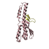
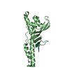
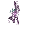
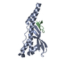
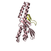
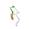
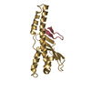
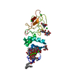
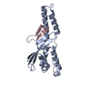
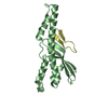
Structure of the SNX32 PX domain in complex with Chlamydial protein IncE in space group I121
Components
IncE
Sorting nexin-32
Keywords
PROTEIN TRANSPORT / PX domain
Function / homology
Function and homology information
retrograde transport, endosome to Golgi / regulation of macroautophagy / phosphatidylinositol binding / protein transport / endosome / cytosol Similarity search - Function
Inclusion membrane protein E / Inclusion membrane protein E / Sorting nexin-5/6/32 / Sorting nexin Vps5-like, C-terminal / Vps5 C terminal like / Phox-like domain / PX Domain / PX domain profile. / PX domain / Phox homology ...Inclusion membrane protein E / Inclusion membrane protein E / Sorting nexin-5/6/32 / Sorting nexin Vps5-like, C-terminal / Vps5 C terminal like / Phox-like domain / PX Domain / PX domain profile. / PX domain / Phox homology / PX domain superfamily / AH/BAR domain superfamily / 2-Layer Sandwich / Alpha Beta Similarity search - Domain/homology
In the structure databanks used in Yorodumi, some data are registered as the other names, "COVID-19 virus" and "2019-nCoV". Here are the details of the virus and the list of structure data.
Jan 31, 2019. EMDB accession codes are about to change! (news from PDBe EMDB page)
EMDB accession codes are about to change! (news from PDBe EMDB page)
The allocation of 4 digits for EMDB accession codes will soon come to an end. Whilst these codes will remain in use, new EMDB accession codes will include an additional digit and will expand incrementally as the available range of codes is exhausted. The current 4-digit format prefixed with “EMD-” (i.e. EMD-XXXX) will advance to a 5-digit format (i.e. EMD-XXXXX), and so on. It is currently estimated that the 4-digit codes will be depleted around Spring 2019, at which point the 5-digit format will come into force.
The EM Navigator/Yorodumi systems omit the EMD- prefix.
Related info.:Q: What is EMD? / ID/Accession-code notation in Yorodumi/EM Navigator
Yorodumi is a browser for structure data from EMDB, PDB, SASBDB, etc.
This page is also the successor to EM Navigator detail page, and also detail information page/front-end page for Omokage search.
The word "yorodu" (or yorozu) is an old Japanese word meaning "ten thousand". "mi" (miru) is to see.
Related info.:EMDB / PDB / SASBDB / Comparison of 3 databanks / Yorodumi Search / Aug 31, 2016. New EM Navigator & Yorodumi / Yorodumi Papers / Jmol/JSmol / Function and homology information / Changes in new EM Navigator and Yorodumi
 Movie
Movie Controller

 Yorodumi
 Open data
 Basic information
 Components
 Keywords
 Function and homology information
 Homo sapiens (human)
 Chlamydia trachomatis (bacteria)
 X-RAY DIFFRACTION /
  Authors
 Australia, 2items
  Citation
 Structure visualization
 Downloads & links
 Other downloads
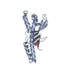


 PDBj
PDBj
 Assembly
 Mass: 18.015 Da / Num. of mol.: 237 / Source method: isolated from a natural source / Formula: H2O
 Sample preparation
 Processing