 Movie
Movie Controller

[English] 日本語
 Yorodumi
- PDB-6e6b: Crystal structure of the Protocadherin GammaB4 extracellular domain -
+ Open data

- Basic information
| Entry | Database: PDB / ID: 6e6b | |||||||||
|---|---|---|---|---|---|---|---|---|---|---|
| Title | Crystal structure of the Protocadherin GammaB4 extracellular domain | |||||||||
 Components Components | Protocadherin gamma B4 | |||||||||
 Keywords Keywords | CELL ADHESION / Cadherin / Polymer / Neuronal self-recognition / Neuronal self-avoidance | |||||||||
| Function / homology |  Function and homology information Function and homology informationhomophilic cell-cell adhesion / cell adhesion / calcium ion binding / membrane / plasma membrane Similarity search - Function | |||||||||
| Biological species |  | |||||||||
| Method |  X-RAY DIFFRACTION / SYNCHROTRON / MOLECULAR REPLACEMENT / Resolution: 4.52 Å X-RAY DIFFRACTION / SYNCHROTRON / MOLECULAR REPLACEMENT / Resolution: 4.52 Å | |||||||||
 Authors Authors | Goodman, K.M. / Mannepalli, S. / Bahna, F. / Honig, B. / Shapiro, L. | |||||||||
| Funding support |  United States, 1items United States, 1items
| |||||||||
 Citation Citation | Journal: Nature / Year: 2019 Title: Visualization of clustered protocadherin neuronal self-recognition complexes. Authors: Julia Brasch / Kerry M Goodman / Alex J Noble / Micah Rapp / Seetha Mannepalli / Fabiana Bahna / Venkata P Dandey / Tristan Bepler / Bonnie Berger / Tom Maniatis / Clinton S Potter / Bridget ...Authors: Julia Brasch / Kerry M Goodman / Alex J Noble / Micah Rapp / Seetha Mannepalli / Fabiana Bahna / Venkata P Dandey / Tristan Bepler / Bonnie Berger / Tom Maniatis / Clinton S Potter / Bridget Carragher / Barry Honig / Lawrence Shapiro / Abstract: Neurite self-recognition and avoidance are fundamental properties of all nervous systems. These processes facilitate dendritic arborization, prevent formation of autapses and allow free interaction ...Neurite self-recognition and avoidance are fundamental properties of all nervous systems. These processes facilitate dendritic arborization, prevent formation of autapses and allow free interaction among non-self neurons. Avoidance among self neurites is mediated by stochastic cell-surface expression of combinations of about 60 isoforms of α-, β- and γ-clustered protocadherin that provide mammalian neurons with single-cell identities. Avoidance is observed between neurons that express identical protocadherin repertoires, and single-isoform differences are sufficient to prevent self-recognition. Protocadherins form isoform-promiscuous cis dimers and isoform-specific homophilic trans dimers. Although these interactions have previously been characterized in isolation, structures of full-length protocadherin ectodomains have not been determined, and how these two interfaces engage in self-recognition between neuronal surfaces remains unknown. Here we determine the molecular arrangement of full-length clustered protocadherin ectodomains in single-isoform self-recognition complexes, using X-ray crystallography and cryo-electron tomography. We determine the crystal structure of the clustered protocadherin γB4 ectodomain, which reveals a zipper-like lattice that is formed by alternating cis and trans interactions. Using cryo-electron tomography, we show that clustered protocadherin γB6 ectodomains tethered to liposomes spontaneously assemble into linear arrays at membrane contact sites, in a configuration that is consistent with the assembly observed in the crystal structure. These linear assemblies pack against each other as parallel arrays to form larger two-dimensional structures between membranes. Our results suggest that the formation of ordered linear assemblies by clustered protocadherins represents the initial self-recognition step in neuronal avoidance, and thus provide support for the isoform-mismatch chain-termination model of protocadherin-mediated self-recognition, which depends on these linear chains. | |||||||||
| History |
|
- Structure visualization
| Structure viewer | Molecule: MolmilJmol/JSmol |
|---|
- Downloads & links
-Download
| PDBx/mmCIF format | 6e6b.cif.gz | 323.1 KB | Display | PDBx/mmCIF format |
|---|---|---|---|---|
| PDB format | pdb6e6b.ent.gz | 210.3 KB | Display | PDB format |
| PDBx/mmJSON format | 6e6b.json.gz | Tree view | PDBx/mmJSON format | |
| Others |  Other downloads Other downloads |
-Validation report
| Arichive directory | https://data.pdbj.org/pub/pdb/validation_reports/e6/6e6bftp://data.pdbj.org/pub/pdb/validation_reports/e6/6e6b | HTTPS FTP |
|---|
-Related structure data
| Related structure data | 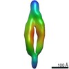 9197C  9198C  9199C  9200C  5t9tS  5v5xS S: Starting model for refinement C: citing same article ( |
|---|---|
| Similar structure data |

-Links
 PDBj
PDBj


- Assembly
| Deposited unit | 
| |||||||||||||||||||||||||||||||||||||||||||||||||||
|---|---|---|---|---|---|---|---|---|---|---|---|---|---|---|---|---|---|---|---|---|---|---|---|---|---|---|---|---|---|---|---|---|---|---|---|---|---|---|---|---|---|---|---|---|---|---|---|---|---|---|---|---|
| 1 |
| |||||||||||||||||||||||||||||||||||||||||||||||||||
| Unit cell |
| |||||||||||||||||||||||||||||||||||||||||||||||||||
| Noncrystallographic symmetry (NCS) | NCS domain:
NCS domain segments: Ens-ID: 1 / Beg auth comp-ID: GLN / Beg label comp-ID: GLN
| |||||||||||||||||||||||||||||||||||||||||||||||||||
| Details | Sedimentation equilibrium analytical ultracentrifugation experiments show that PcdhgB4 EC1-6 is a dimer-of-dimers in solution. However in both the crystal structure and cryo-electron tomography of PcdhgB4 on membrane reveals a one-dimensional lattice of alternating C-terminal (cis) and N-terminal (trans) interactions, the same as those used to form the dimer-of-dimers observed in solution but generating a polymer rather than a discrete dimer-of-dimers. |
-Components
-Protein / Non-polymers , 2 types, 32 molecules AB
| #1: Protein | Mass: 70991.781 Da / Num. of mol.: 2 Source method: isolated from a genetically manipulated source Details: Protocadherin extracellular cadherin domains 1-6 with C-terminal octahistidine expression tag Source: (gene. exp.)  Homo sapiens (human) / References: UniProt: Q91XX6 Homo sapiens (human) / References: UniProt: Q91XX6#5: Chemical | ChemComp-CA /  Mass: 40.078 Da / Num. of mol.: 30 / Source method: obtained synthetically / Formula: Ca Mass: 40.078 Da / Num. of mol.: 30 / Source method: obtained synthetically / Formula: Ca |
|---|
-Sugars , 5 types, 10 molecules 

| #2: Polysaccharide | 2-acetamido-2-deoxy-beta-D-glucopyranose-(1-4)-2-acetamido-2-deoxy-beta-D-glucopyranose Source method: isolated from a genetically manipulated source | ||
|---|---|---|---|
| #3: Polysaccharide | alpha-D-mannopyranose-(1-3)-[alpha-D-mannopyranose-(1-6)]beta-D-mannopyranose-(1-4)-2-acetamido-2- ...alpha-D-mannopyranose-(1-3)-[alpha-D-mannopyranose-(1-6)]beta-D-mannopyranose-(1-4)-2-acetamido-2-deoxy-beta-D-glucopyranose-(1-4)-[alpha-L-fucopyranose-(1-6)]2-acetamido-2-deoxy-beta-D-glucopyranose Source method: isolated from a genetically manipulated source | ||
| #4: Polysaccharide | 2-acetamido-2-deoxy-beta-D-glucopyranose-(1-4)-[alpha-L-fucopyranose-(1-6)]2-acetamido-2-deoxy-beta- ...2-acetamido-2-deoxy-beta-D-glucopyranose-(1-4)-[alpha-L-fucopyranose-(1-6)]2-acetamido-2-deoxy-beta-D-glucopyranose Source method: isolated from a genetically manipulated source | ||
| #6: Sugar | ChemComp-MAN /  Type: D-saccharide, alpha linking / Mass: 180.156 Da / Num. of mol.: 4 Type: D-saccharide, alpha linking / Mass: 180.156 Da / Num. of mol.: 4Source method: isolated from a genetically manipulated source Formula: C6H12O6 #7: Sugar |  Type: D-saccharide, beta linking / Mass: 221.208 Da / Num. of mol.: 3 Type: D-saccharide, beta linking / Mass: 221.208 Da / Num. of mol.: 3Source method: isolated from a genetically manipulated source Formula: C8H15NO6 |
-Details
| Has protein modification | Y |
|---|
-Experimental details
-Experiment
| Experiment | Method: X-RAY DIFFRACTION / Number of used crystals: 1 |
|---|
- Sample preparation
| Crystal | Density Matthews: 5.55 Å3/Da / Density % sol: 77.83 % |
|---|---|
| Crystal grow | Temperature: 295 K / Method: batch mode / pH: 7.5 Details: 10% (w/v) PEG8000, 20% ethylene glycol, 10% Morpheus Amino Acids (Molecular Dimensions), and 0.1 M Morpheus Buffer System 2 (Hepes/MOPS buffer; Molecular Dimensions) pH 7.5 |
-Data collection
| Diffraction | Mean temperature: 100 K / Serial crystal experiment: N |
|---|---|
| Diffraction source | Source: SYNCHROTRON / Site: APS / Beamline: 24-ID-C / Wavelength: 0.97919 Å |
| Detector | Type: DECTRIS PILATUS 6M-F / Detector: PIXEL / Date: Dec 1, 2016 |
| Radiation | Protocol: SINGLE WAVELENGTH / Monochromatic (M) / Laue (L): M / Scattering type: x-ray |
| Radiation wavelength | Wavelength: 0.97919 Å / Relative weight: 1 |
| Reflection | Resolution: 4.5→40 Å / Num. obs: 8694 / % possible obs: 93.4 % / Redundancy: 2.8 % / Biso Wilson estimate: 66.09 Å2 / CC1/2: 0.995 / Rmerge(I) obs: 0.113 / Rpim(I) all: 0.078 / Rrim(I) all: 0.138 / Net I/σ(I): 5.2 |
| Reflection shell | Resolution: 4.5→5.05 Å / Redundancy: 3 % / Rmerge(I) obs: 0.173 / Mean I/σ(I) obs: 5.7 / Num. unique obs: 317 / CC1/2: 0.973 / Rpim(I) all: 0.119 / Rrim(I) all: 0.211 |
- Processing
| Software |
| ||||||||||||||||||||||||||||
|---|---|---|---|---|---|---|---|---|---|---|---|---|---|---|---|---|---|---|---|---|---|---|---|---|---|---|---|---|---|
| Refinement | Method to determine structure: MOLECULAR REPLACEMENT Starting model: 5V5X, 5T9T Resolution: 4.52→38.29 Å / SU ML: 0.7857 / Cross valid method: FREE R-VALUE / σ(F): 1.35 / Phase error: 34.4867 Details: Due to the severe anisotropy of the data ellipsoidal resolution limits of 6.8, 6.0, and 4.5 angstroms were applied along the three principal axes. The completeness with respect to a sphere ...Details: Due to the severe anisotropy of the data ellipsoidal resolution limits of 6.8, 6.0, and 4.5 angstroms were applied along the three principal axes. The completeness with respect to a sphere at 4.5 angstroms is therefore very low (46.9%), however the completeness within the ellipsoidal limits is 93.4%.
| ||||||||||||||||||||||||||||
| Solvent computation | Shrinkage radii: 0.9 Å / VDW probe radii: 1.11 Å | ||||||||||||||||||||||||||||
| Displacement parameters | Biso mean: 138.7 Å2 | ||||||||||||||||||||||||||||
| Refinement step | Cycle: LAST / Resolution: 4.52→38.29 Å
| ||||||||||||||||||||||||||||
| Refine LS restraints |
| ||||||||||||||||||||||||||||
| LS refinement shell |
|
