 Movie
Movie Controller


+ Open data

- Basic information
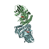
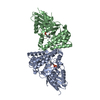
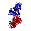
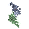
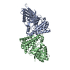
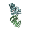
| Entry | Database: PDB / ID: 6e2u | ||||||
|---|---|---|---|---|---|---|---|
| Title | MDDEF in complex with MVAPP, AMPPCP and Magnesium | ||||||
 Components Components | Mevalonate diphosphate decarboxylase | ||||||
 Keywords Keywords | LYASE / Mevalonate diphosphate decarboxylase | ||||||
| Function / homology |  Function and homology information Function and homology informationdiphosphomevalonate decarboxylase / diphosphomevalonate decarboxylase activity / isopentenyl diphosphate biosynthetic process, mevalonate pathway / ATP binding / metal ion binding / cytosol Similarity search - Function | ||||||
| Biological species |   Enterococcus faecalis (bacteria) Enterococcus faecalis (bacteria) | ||||||
| Method |  X-RAY DIFFRACTION / MOLECULAR REPLACEMENT / Resolution: 2.05 Å X-RAY DIFFRACTION / MOLECULAR REPLACEMENT / Resolution: 2.05 Å | ||||||
 Authors Authors | Stauffacher, C.V. / Chen, C.-L. | ||||||
 Citation Citation | Journal: Nat Commun / Year: 2020 Title: Visualizing the enzyme mechanism of mevalonate diphosphate decarboxylase. Authors: Chen, C.L. / Paul, L.N. / Mermoud, J.C. / Steussy, C.N. / Stauffacher, C.V. | ||||||
| History |
|
- Structure visualization
| Structure viewer | Molecule: MolmilJmol/JSmol |
|---|
- Downloads & links
-Download
| PDBx/mmCIF format | 6e2u.cif.gz | 137.6 KB | Display | PDBx/mmCIF format |
|---|---|---|---|---|
| PDB format | pdb6e2u.ent.gz | 104.9 KB | Display | PDB format |
| PDBx/mmJSON format | 6e2u.json.gz | Tree view | PDBx/mmJSON format | |
| Others |  Other downloads Other downloads |
-Validation report
| Arichive directory | https://data.pdbj.org/pub/pdb/validation_reports/e2/6e2uftp://data.pdbj.org/pub/pdb/validation_reports/e2/6e2u | HTTPS FTP |
|---|
-Related structure data
| Related structure data | 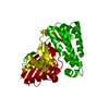 6e2sC  6e2tC  6e2vC  6e2wC  6e2yC 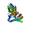 5v2lS S: Starting model for refinement C: citing same article ( |
|---|---|
| Similar structure data |


-Links
 PDBj
PDBj


- Assembly
| Deposited unit | 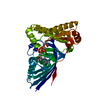
| ||||||||
|---|---|---|---|---|---|---|---|---|---|
| 1 | 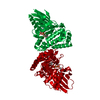
| ||||||||
| Unit cell |
|
-Components
| #1: Protein | Mass: 39276.363 Da / Num. of mol.: 1 Source method: isolated from a genetically manipulated source Source: (gene. exp.) Enterococcus faecalis (bacteria) / Gene: mvaD, B6S42_02310 / Production host: References: UniProt: Q9FD68, diphosphomevalonate decarboxylase |
|---|---|
| #2: Chemical | ChemComp-DP6 / (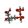  Mass: 308.117 Da / Num. of mol.: 1 / Source method: obtained synthetically / Formula: C6H14O10P2 Mass: 308.117 Da / Num. of mol.: 1 / Source method: obtained synthetically / Formula: C6H14O10P2 |
| #3: Chemical | ChemComp-ACP /   Mass: 505.208 Da / Num. of mol.: 1 / Source method: obtained synthetically / Formula: C11H18N5O12P3 / Comment: AMP-PCP, energy-carrying molecule analogue*YM Mass: 505.208 Da / Num. of mol.: 1 / Source method: obtained synthetically / Formula: C11H18N5O12P3 / Comment: AMP-PCP, energy-carrying molecule analogue*YM |
| #4: Chemical | ChemComp-MG /   Mass: 24.305 Da / Num. of mol.: 1 / Source method: obtained synthetically / Formula: Mg Mass: 24.305 Da / Num. of mol.: 1 / Source method: obtained synthetically / Formula: Mg |
| #5: Water | ChemComp-HOH /  Mass: 18.015 Da / Num. of mol.: 90 / Source method: isolated from a natural source / Formula: H2O Mass: 18.015 Da / Num. of mol.: 90 / Source method: isolated from a natural source / Formula: H2O |
-Experimental details
-Experiment
| Experiment | Method: X-RAY DIFFRACTION / Number of used crystals: 1 |
|---|
- Sample preparation
| Crystal | Density Matthews: 2.27 Å3/Da / Density % sol: 45.93 % |
|---|---|
| Crystal grow | Temperature: 293 K / Method: vapor diffusion, sitting drop / pH: 4.6 Details: Crystals were produced under the conditions of 1.6 M ammonium sulfate, 50 mM sodium acetate, pH 4.6; buffer exchange was performed under the conditions of 26 % PEG 3350, 5 mM MgCl2 and 50 mM ...Details: Crystals were produced under the conditions of 1.6 M ammonium sulfate, 50 mM sodium acetate, pH 4.6; buffer exchange was performed under the conditions of 26 % PEG 3350, 5 mM MgCl2 and 50 mM sodium acetate, pH 4.6; Crystals were soaked with MVAPP, followed by AMPPCP. Then condensation was conducted at 30 % PEG 3350, 15% PEG 400, 5 mM MgCl2, 50 mM sodium acetate, pH 4.6 |
-Data collection
| Diffraction | Mean temperature: 100 K |
|---|---|
| Diffraction source | Source: ROTATING ANODE / Type: RIGAKU RUH2R / Wavelength: 1.54 Å |
| Detector | Type: RIGAKU RAXIS IV++ / Detector: IMAGE PLATE / Date: Oct 14, 2014 |
| Radiation | Protocol: SINGLE WAVELENGTH / Monochromatic (M) / Laue (L): M / Scattering type: x-ray |
| Radiation wavelength | Wavelength: 1.54 Å / Relative weight: 1 |
| Reflection | Resolution: 2.045→30 Å / Num. obs: 23332 / % possible obs: 96.6 % / Redundancy: 5.1 % / Net I/σ(I): 38.6 |
| Reflection shell | Resolution: 2.05→2.12 Å / Redundancy: 4.9 % / Mean I/σ(I) obs: 2.5 / Num. unique obs: 2246 / % possible all: 98.5 |
- Processing
| Software |
| |||||||||||||||||||||||||||||||||||||||||||||||||||||||||||||||
|---|---|---|---|---|---|---|---|---|---|---|---|---|---|---|---|---|---|---|---|---|---|---|---|---|---|---|---|---|---|---|---|---|---|---|---|---|---|---|---|---|---|---|---|---|---|---|---|---|---|---|---|---|---|---|---|---|---|---|---|---|---|---|---|---|
| Refinement | Method to determine structure: MOLECULAR REPLACEMENT Starting model: 5V2L Resolution: 2.05→28.83 Å / SU ML: 0.19 / Cross valid method: FREE R-VALUE / σ(F): 1.34 / Phase error: 19.98
| |||||||||||||||||||||||||||||||||||||||||||||||||||||||||||||||
| Solvent computation | Shrinkage radii: 0.9 Å / VDW probe radii: 1.11 Å | |||||||||||||||||||||||||||||||||||||||||||||||||||||||||||||||
| Refinement step | Cycle: LAST / Resolution: 2.05→28.83 Å
| |||||||||||||||||||||||||||||||||||||||||||||||||||||||||||||||
| Refine LS restraints |
| |||||||||||||||||||||||||||||||||||||||||||||||||||||||||||||||
| LS refinement shell |
|
