 Movie
Movie Controller

[English] 日本語
 Yorodumi
- PDB-6dyn: C-terminal condensation domain of Ebony in complex with Histamine -
+ Open data

- Basic information
| Entry | Database: PDB / ID: 6dyn | ||||||
|---|---|---|---|---|---|---|---|
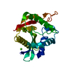
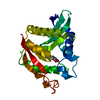
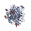
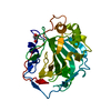
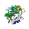
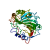
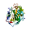
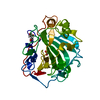
| Title | C-terminal condensation domain of Ebony in complex with Histamine | ||||||
 Components Components | Ebony | ||||||
 Keywords Keywords | BIOSYNTHETIC PROTEIN / NRPS / Condensation domain / Peptide-bond formation | ||||||
| Function / homology |  Function and homology information Function and homology informationANL, N-terminal domain / AMP-binding, conserved site / Putative AMP-binding domain signature. / AMP-dependent synthetase/ligase / AMP-binding enzyme / AMP-binding enzyme, C-terminal domain superfamily / Phosphopantetheine attachment site / ACP-like superfamily / Carrier protein (CP) domain profile. / Phosphopantetheine binding ACP domain Similarity search - Domain/homology | ||||||
| Biological species |  | ||||||
| Method |  X-RAY DIFFRACTION / SYNCHROTRON / MOLECULAR REPLACEMENT / Resolution: 2.102 Å X-RAY DIFFRACTION / SYNCHROTRON / MOLECULAR REPLACEMENT / Resolution: 2.102 Å | ||||||
 Authors Authors | Izore, T. / Tailhades, J. / Hansen, M.H. / Kaczmarski, J.A. / Jackson, C.J. / Cryle, M.J. | ||||||
| Funding support |  Australia, 1items Australia, 1items
| ||||||
 Citation Citation | Journal: Proc. Natl. Acad. Sci. U.S.A. / Year: 2019 Title: Drosophila melanogasternonribosomal peptide synthetase Ebony encodes an atypical condensation domain. Authors: Izore, T. / Tailhades, J. / Hansen, M.H. / Kaczmarski, J.A. / Jackson, C.J. / Cryle, M.J. | ||||||
| History |
|
- Structure visualization
| Structure viewer | Molecule: MolmilJmol/JSmol |
|---|
- Downloads & links
-Download
| PDBx/mmCIF format | 6dyn.cif.gz | 63.6 KB | Display | PDBx/mmCIF format |
|---|---|---|---|---|
| PDB format | pdb6dyn.ent.gz | 43.3 KB | Display | PDB format |
| PDBx/mmJSON format | 6dyn.json.gz | Tree view | PDBx/mmJSON format | |
| Others |  Other downloads Other downloads |
-Validation report
| Arichive directory | https://data.pdbj.org/pub/pdb/validation_reports/dy/6dynftp://data.pdbj.org/pub/pdb/validation_reports/dy/6dyn | HTTPS FTP |
|---|
-Related structure data
| Related structure data |  6dymSC  6dyoC 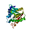 6dyrC 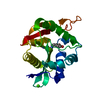 6dysC S: Starting model for refinement C: citing same article ( |
|---|---|
| Similar structure data |

-Links
 PDBj
PDBj

- Assembly
| Deposited unit | 
| ||||||||||
|---|---|---|---|---|---|---|---|---|---|---|---|
| 1 |
| ||||||||||
| Unit cell |
|
-Components
| #1: Protein | Mass: 25991.506 Da / Num. of mol.: 1 Source method: isolated from a genetically manipulated source Source: (gene. exp.)  |
|---|---|
| #2: Chemical | ChemComp-HSM /   Mass: 111.145 Da / Num. of mol.: 1 / Source method: obtained synthetically / Formula: C5H9N3 Mass: 111.145 Da / Num. of mol.: 1 / Source method: obtained synthetically / Formula: C5H9N3 |
| #3: Chemical | ChemComp-CA /   Mass: 40.078 Da / Num. of mol.: 1 / Source method: obtained synthetically / Formula: Ca Mass: 40.078 Da / Num. of mol.: 1 / Source method: obtained synthetically / Formula: Ca |
| #4: Water | ChemComp-HOH /  Mass: 18.015 Da / Num. of mol.: 146 / Source method: isolated from a natural source / Formula: H2O Mass: 18.015 Da / Num. of mol.: 146 / Source method: isolated from a natural source / Formula: H2O |
-Experimental details
-Experiment
| Experiment | Method: X-RAY DIFFRACTION / Number of used crystals: 1 |
|---|
- Sample preparation
| Crystal | Density Matthews: 2.38 Å3/Da / Density % sol: 48.24 % / Description: Long thin needles |
|---|---|
| Crystal grow | Temperature: 293 K / Method: vapor diffusion, sitting drop / pH: 7.5 / Details: HEPES, PEG 400, CaCl2 |
-Data collection
| Diffraction | Mean temperature: 100 K / Serial crystal experiment: N |
|---|---|
| Diffraction source | Source: SYNCHROTRON / Site: Australian Synchrotron / Beamline: MX1 / Wavelength: 0.9537 Å |
| Detector | Type: ADSC QUANTUM 210r / Detector: CCD / Date: Feb 8, 2018 |
| Radiation | Protocol: SINGLE WAVELENGTH / Monochromatic (M) / Laue (L): M / Scattering type: x-ray |
| Radiation wavelength | Wavelength: 0.9537 Å / Relative weight: 1 |
| Reflection | Resolution: 2.1→40.97 Å / Num. obs: 13655 / % possible obs: 99.6 % / Redundancy: 4.4 % / Biso Wilson estimate: 27.6 Å2 / Rpim(I) all: 0.05 / Net I/av σ(I): 11.9 / Net I/σ(I): 11.9 |
| Reflection shell | Resolution: 2.1→2.16 Å / Mean I/σ(I) obs: 2 / CC1/2: 0.63 / Rpim(I) all: 0.4 |
- Processing
| Software |
| ||||||||||||||||
|---|---|---|---|---|---|---|---|---|---|---|---|---|---|---|---|---|---|
| Refinement | Method to determine structure: MOLECULAR REPLACEMENT Starting model: 6DYM Resolution: 2.102→40.97 Å / Cross valid method: FREE R-VALUE
| ||||||||||||||||
| Refinement step | Cycle: LAST / Resolution: 2.102→40.97 Å
|
