 Movie
Movie Controller

[English] 日本語
 Yorodumi
- PDB-6drq: The crystal structure of SatS c-terminal domain in complex with b... -
+ Open data

- Basic information
| Entry | Database: PDB / ID: 6drq | ||||||
|---|---|---|---|---|---|---|---|
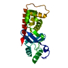
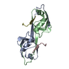
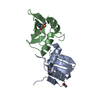
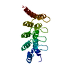
| Title | The crystal structure of SatS c-terminal domain in complex with bromine | ||||||
 Components Components | Primosomal protein | ||||||
 Keywords Keywords | CHAPERONE / SecA2 / protein export / Structural Genomics / PSI-2 / Protein Structure Initiative / TB Structural Genomics Consortium / TBSGC | ||||||
| Function / homology | BROMIDE ION / :  Function and homology information Function and homology information | ||||||
| Biological species |   Mycobacterium tuberculosis (bacteria) Mycobacterium tuberculosis (bacteria) | ||||||
| Method |  X-RAY DIFFRACTION / SYNCHROTRON / SAD / Resolution: 2.3 Å X-RAY DIFFRACTION / SYNCHROTRON / SAD / Resolution: 2.3 Å | ||||||
 Authors Authors | Hughes, R.C. / Sacchettini, J.C. / TB Structural Genomics Consortium (TBSGC) | ||||||
| Funding support |  United States, 1items United States, 1items
| ||||||
 Citation Citation | Journal: Elife / Year: 2019 Title: Mycobacterium tuberculosisSatS is a chaperone for the SecA2 protein export pathway. Authors: Miller, B.K. / Hughes, R. / Ligon, L.S. / Rigel, N.W. / Malik, S. / Anjuwon-Foster, B.R. / Sacchettini, J.C. / Braunstein, M. | ||||||
| History |
|
- Structure visualization
| Structure viewer | Molecule: MolmilJmol/JSmol |
|---|
- Downloads & links
-Download
| PDBx/mmCIF format | 6drq.cif.gz | 49.2 KB | Display | PDBx/mmCIF format |
|---|---|---|---|---|
| PDB format | pdb6drq.ent.gz | 33.5 KB | Display | PDB format |
| PDBx/mmJSON format | 6drq.json.gz | Tree view | PDBx/mmJSON format | |
| Others |  Other downloads Other downloads |
-Validation report
| Arichive directory | https://data.pdbj.org/pub/pdb/validation_reports/dr/6drqftp://data.pdbj.org/pub/pdb/validation_reports/dr/6drq | HTTPS FTP |
|---|
-Related structure data


-Links
 PDBj
PDBj


- Assembly
| Deposited unit | 
| ||||||||
|---|---|---|---|---|---|---|---|---|---|
| 1 |
| ||||||||
| Unit cell |
|
-Components
| #1: Protein | Mass: 20620.805 Da / Num. of mol.: 1 Source method: isolated from a genetically manipulated source Source: (gene. exp.) Mycobacterium tuberculosis (bacteria) / Strain: ATCC 25618 / H37Rv / Gene: LH57_18085 / Production host: | ||
|---|---|---|---|
| #2: Chemical |   Mass: 79.904 Da / Num. of mol.: 3 / Source method: obtained synthetically / Formula: Br Mass: 79.904 Da / Num. of mol.: 3 / Source method: obtained synthetically / Formula: Br#3: Water | ChemComp-HOH / |  Mass: 18.015 Da / Num. of mol.: 13 / Source method: isolated from a natural source / Formula: H2O Mass: 18.015 Da / Num. of mol.: 13 / Source method: isolated from a natural source / Formula: H2O |
-Experimental details
-Experiment
| Experiment | Method: X-RAY DIFFRACTION / Number of used crystals: 1 |
|---|
- Sample preparation
| Crystal | Density Matthews: 2.28 Å3/Da / Density % sol: 45.98 % |
|---|---|
| Crystal grow | Temperature: 289 K / Method: vapor diffusion, sitting drop / Details: 3.5M ammonium citrate |
-Data collection
| Diffraction | Mean temperature: 129 K |
|---|---|
| Diffraction source | Source: SYNCHROTRON / Site: APS / Beamline: 23-ID-B / Wavelength: 0.902 Å |
| Detector | Type: DECTRIS PILATUS3 6M / Detector: PIXEL / Date: Mar 26, 2015 |
| Radiation | Protocol: SINGLE WAVELENGTH / Monochromatic (M) / Laue (L): M / Scattering type: x-ray |
| Radiation wavelength | Wavelength: 0.902 Å / Relative weight: 1 |
| Reflection | Resolution: 2.3→99 Å / Num. obs: 8721 / % possible obs: 99.2 % / Redundancy: 3.8 % / Rmerge(I) obs: 0.05 / Rsym value: 0.053 / Net I/σ(I): 27.4 |
| Reflection shell | Resolution: 2.3→2.34 Å / Rmerge(I) obs: 0.105 / Num. unique all: 806 |
- Processing
| Software |
| ||||||||||||||||||||||||||||
|---|---|---|---|---|---|---|---|---|---|---|---|---|---|---|---|---|---|---|---|---|---|---|---|---|---|---|---|---|---|
| Refinement | Method to determine structure: SAD / Resolution: 2.3→42.129 Å / SU ML: 0.25 / Cross valid method: FREE R-VALUE / σ(F): 1.36 / Phase error: 28.32
| ||||||||||||||||||||||||||||
| Solvent computation | Shrinkage radii: 0.9 Å / VDW probe radii: 1.11 Å | ||||||||||||||||||||||||||||
| Refinement step | Cycle: LAST / Resolution: 2.3→42.129 Å
| ||||||||||||||||||||||||||||
| Refine LS restraints |
| ||||||||||||||||||||||||||||
| LS refinement shell |
|
