 Movie
Movie Controller

[English] 日本語
 Yorodumi
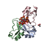
- PDB-6dkl: Crystal Structure of a Rationally Designed Six-Fold Symmetric DNA... -
+ Open data

- Basic information
| Entry | Database: PDB / ID: 6dkl | |||||||||
|---|---|---|---|---|---|---|---|---|---|---|
| Title | Crystal Structure of a Rationally Designed Six-Fold Symmetric DNA Scaffold | |||||||||
 Components Components |
| |||||||||
 Keywords Keywords | DNA / DNA Nanotechnology / Structural DNA Nanotechnology / self-assembly / crystal design / DNA crystals | |||||||||
| Function / homology | DNA / DNA (> 10) Function and homology information Function and homology information | |||||||||
| Biological species | synthetic construct (others) | |||||||||
| Method |  X-RAY DIFFRACTION / SYNCHROTRON / SAD / Resolution: 3.034 Å X-RAY DIFFRACTION / SYNCHROTRON / SAD / Resolution: 3.034 Å | |||||||||
 Authors Authors | Simmons, C.R. / Zhang, F. / Yan, H. | |||||||||
| Funding support |  United States, 2items United States, 2items
| |||||||||
 Citation Citation | Journal: Angew. Chem. Int. Ed. Engl. / Year: 2018 Title: Self-Assembly of a 3D DNA Crystal Structure with Rationally Designed Six-Fold Symmetry. Authors: Zhang, F. / Simmons, C.R. / Gates, J. / Liu, Y. / Yan, H. | |||||||||
| History |
|
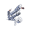
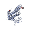
- Structure visualization
| Structure viewer | Molecule: MolmilJmol/JSmol |
|---|
- Downloads & links
-Download
| PDBx/mmCIF format | 6dkl.cif.gz | 42.8 KB | Display | PDBx/mmCIF format |
|---|---|---|---|---|
| PDB format | pdb6dkl.ent.gz | 30.7 KB | Display | PDB format |
| PDBx/mmJSON format | 6dkl.json.gz | Tree view | PDBx/mmJSON format | |
| Others |  Other downloads Other downloads |
-Validation report
| Summary document | 6dkl_validation.pdf.gz | 386.5 KB | Display | wwPDB validaton report |
|---|---|---|---|---|
| Full document | 6dkl_full_validation.pdf.gz | 387 KB | Display | |
| Data in XML | 6dkl_validation.xml.gz | 3.1 KB | Display | |
| Data in CIF | 6dkl_validation.cif.gz | 3.7 KB | Display | |
| Arichive directory | https://data.pdbj.org/pub/pdb/validation_reports/dk/6dklftp://data.pdbj.org/pub/pdb/validation_reports/dk/6dkl | HTTPS FTP |
-Related structure data
| Similar structure data |
|---|


-Links
 PDBj
PDBj


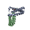
- Assembly
| Deposited unit | 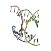
| ||||||||
|---|---|---|---|---|---|---|---|---|---|
| 1 |
| ||||||||
| Unit cell |
|
-Components
| #1: DNA chain | Mass: 2764.837 Da / Num. of mol.: 1 / Source method: obtained synthetically / Source: (synth.) synthetic construct (others) |
|---|---|
| #2: DNA chain | Mass: 2973.971 Da / Num. of mol.: 1 / Source method: obtained synthetically / Source: (synth.) synthetic construct (others) |
| #3: DNA chain | Mass: 3371.193 Da / Num. of mol.: 1 / Source method: obtained synthetically / Source: (synth.) synthetic construct (others) |
| #4: DNA chain | Mass: 3687.417 Da / Num. of mol.: 1 / Source method: obtained synthetically / Source: (synth.) synthetic construct (others) |
-Experimental details
-Experiment
| Experiment | Method: X-RAY DIFFRACTION / Number of used crystals: 1 |
|---|
- Sample preparation
| Crystal grow | Temperature: 298 K / Method: vapor diffusion, sitting drop Details: 50 mM Cacodylate pH 6.0, 10 mM MgCl2, 200 mM sodium citrate, 5% isopropanol Temp details: Temperature gradient was generated from 60C-25C for .3 degrees per hour. |
|---|
-Data collection
| Diffraction | Mean temperature: 100 K |
|---|---|
| Diffraction source | Source: SYNCHROTRON / Site: APS / Beamline: 19-ID / Wavelength: 1 Å |
| Detector | Type: DECTRIS PILATUS 6M / Detector: PIXEL / Date: Oct 21, 2017 |
| Radiation | Monochromator: Si(111) / Protocol: SINGLE WAVELENGTH / Monochromatic (M) / Laue (L): M / Scattering type: x-ray |
| Radiation wavelength | Wavelength: 1 Å / Relative weight: 1 |
| Reflection | Resolution: 3.034→50 Å / Num. obs: 6788 / % possible obs: 91.8 % / Redundancy: 6.8 % / CC1/2: 0.98 / Rmerge(I) obs: 0.106 / Rpim(I) all: 0.044 / Rrim(I) all: 0.115 / Net I/σ(I): 42.1 |
| Reflection shell | Resolution: 3.034→3.1 Å / Num. unique obs: 252 / CC1/2: 0.93 / Rpim(I) all: 0.2 |
- Processing
| Software |
| ||||||||||||||||||||||||||||||||||||||||||
|---|---|---|---|---|---|---|---|---|---|---|---|---|---|---|---|---|---|---|---|---|---|---|---|---|---|---|---|---|---|---|---|---|---|---|---|---|---|---|---|---|---|---|---|
| Refinement | Method to determine structure: SAD / Resolution: 3.034→37.1758 Å / SU ML: 0.29 / Cross valid method: FREE R-VALUE / σ(F): 1.42 / Phase error: 41.42
| ||||||||||||||||||||||||||||||||||||||||||
| Solvent computation | Shrinkage radii: 0.9 Å / VDW probe radii: 1.11 Å | ||||||||||||||||||||||||||||||||||||||||||
| Refinement step | Cycle: LAST / Resolution: 3.034→37.1758 Å
| ||||||||||||||||||||||||||||||||||||||||||
| Refine LS restraints |
| ||||||||||||||||||||||||||||||||||||||||||
| LS refinement shell |
|
