 Movie
Movie Controller


+ Open data

- Basic information
| Entry | Database: PDB / ID: 6d6w | |||||||||
|---|---|---|---|---|---|---|---|---|---|---|
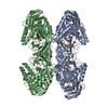
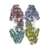
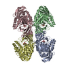
| Title | Bacteroides uniformis beta-glucuronidase 1 bound to glucuronate | |||||||||
 Components Components | Beta-galactosidase/beta-glucuronidase | |||||||||
 Keywords Keywords | HYDROLASE / glycosyl hydrolase / beta-glucuronidase | |||||||||
| Function / homology |  Function and homology information Function and homology informationbeta-glucuronidase / beta-glucuronidase activity / carbohydrate metabolic process Similarity search - Function | |||||||||
| Biological species |  Bacteroides uniformis (bacteria) Bacteroides uniformis (bacteria) | |||||||||
| Method |  X-RAY DIFFRACTION / SYNCHROTRON / MOLECULAR REPLACEMENT / Resolution: 1.8 Å X-RAY DIFFRACTION / SYNCHROTRON / MOLECULAR REPLACEMENT / Resolution: 1.8 Å | |||||||||
 Authors Authors | Walton, W.G. / Pellock, S.J. / Redinbo, M.R. | |||||||||
| Funding support |  United States, 2items United States, 2items
| |||||||||
 Citation Citation | Journal: J. Biol. Chem. / Year: 2018 Title: Three structurally and functionally distinct beta-glucuronidases from the human gut microbeBacteroides uniformis. Authors: Pellock, S.J. / Walton, W.G. / Biernat, K.A. / Torres-Rivera, D. / Creekmore, B.C. / Xu, Y. / Liu, J. / Tripathy, A. / Stewart, L.J. / Redinbo, M.R. | |||||||||
| History |
|
- Structure visualization
| Structure viewer | Molecule: MolmilJmol/JSmol |
|---|
- Downloads & links
-Download
| PDBx/mmCIF format | 6d6w.cif.gz | 591.1 KB | Display | PDBx/mmCIF format |
|---|---|---|---|---|
| PDB format | pdb6d6w.ent.gz | 470.3 KB | Display | PDB format |
| PDBx/mmJSON format | 6d6w.json.gz | Tree view | PDBx/mmJSON format | |
| Others |  Other downloads Other downloads |
-Validation report
| Arichive directory | https://data.pdbj.org/pub/pdb/validation_reports/d6/6d6wftp://data.pdbj.org/pub/pdb/validation_reports/d6/6d6w | HTTPS FTP |
|---|
-Related structure data
| Related structure data | 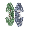 6d1nC  6d1pC  6d41C  6d50C  6d7fC  6d89C  6d8gC  6d8kC C: citing same article ( |
|---|---|
| Similar structure data |


-Links
 PDBj
PDBj

- Assembly
| Deposited unit | 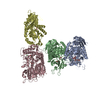
| |||||||||||||||||||||||||||
|---|---|---|---|---|---|---|---|---|---|---|---|---|---|---|---|---|---|---|---|---|---|---|---|---|---|---|---|---|
| 1 | 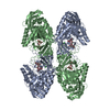
| |||||||||||||||||||||||||||
| 2 | 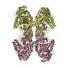
| |||||||||||||||||||||||||||
| 3 | 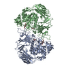
| |||||||||||||||||||||||||||
| 4 | 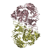
| |||||||||||||||||||||||||||
| Unit cell |
| |||||||||||||||||||||||||||
| Components on special symmetry positions |
|
-Components
| #1: Protein | Mass: 70098.500 Da / Num. of mol.: 4 Source method: isolated from a genetically manipulated source Source: (gene. exp.) Bacteroides uniformis (bacteria) / Gene: uidA_4, ERS417307_01040 / Production host: #2: Sugar | ChemComp-GCU /   Type: D-saccharide, alpha linking / Mass: 194.139 Da / Num. of mol.: 4 / Source method: obtained synthetically / Formula: C6H10O7 / Feature type: SUBJECT OF INVESTIGATION Type: D-saccharide, alpha linking / Mass: 194.139 Da / Num. of mol.: 4 / Source method: obtained synthetically / Formula: C6H10O7 / Feature type: SUBJECT OF INVESTIGATION#3: Chemical | ChemComp-GOL /   Mass: 92.094 Da / Num. of mol.: 12 / Source method: obtained synthetically / Formula: C3H8O3 Mass: 92.094 Da / Num. of mol.: 12 / Source method: obtained synthetically / Formula: C3H8O3#4: Chemical | ChemComp-CL /   Mass: 35.453 Da / Num. of mol.: 4 / Source method: obtained synthetically / Formula: Cl Mass: 35.453 Da / Num. of mol.: 4 / Source method: obtained synthetically / Formula: Cl#5: Water | ChemComp-HOH / |  Mass: 18.015 Da / Num. of mol.: 4262 / Source method: isolated from a natural source / Formula: H2O Mass: 18.015 Da / Num. of mol.: 4262 / Source method: isolated from a natural source / Formula: H2O |
|---|
-Experimental details
-Experiment
| Experiment | Method: X-RAY DIFFRACTION / Number of used crystals: 1 |
|---|
- Sample preparation
| Crystal | Density Matthews: 2.58 Å3/Da / Density % sol: 52.25 % |
|---|---|
| Crystal grow | Temperature: 293.15 K / Method: vapor diffusion, hanging drop Details: 0.1M Sodium Citrate: Citrate Acid, pH 5.5, 20% PEG 3000, 20mM Glucuronic Acid |
-Data collection
| Diffraction | Mean temperature: 100 K |
|---|---|
| Diffraction source | Source: SYNCHROTRON / Site: APS / Beamline: 23-ID-D / Wavelength: 1.03319 Å |
| Detector | Type: DECTRIS PILATUS3 6M / Detector: PIXEL / Date: Dec 1, 2017 |
| Radiation | Protocol: SINGLE WAVELENGTH / Monochromatic (M) / Laue (L): M / Scattering type: x-ray |
| Radiation wavelength | Wavelength: 1.03319 Å / Relative weight: 1 |
| Reflection | Resolution: 1.8→29.98 Å / Num. all: 261960 / Num. obs: 261936 / % possible obs: 100 % / Redundancy: 6.7 % / Net I/σ(I): 11.8 |
| Reflection shell | Resolution: 1.8→1.83 Å |
- Processing
| Software |
| |||||||||||||||||||||||||||||||||||||||||||||||||||||||||||||||||||||||||||||||||||||||||||||||||||||||||
|---|---|---|---|---|---|---|---|---|---|---|---|---|---|---|---|---|---|---|---|---|---|---|---|---|---|---|---|---|---|---|---|---|---|---|---|---|---|---|---|---|---|---|---|---|---|---|---|---|---|---|---|---|---|---|---|---|---|---|---|---|---|---|---|---|---|---|---|---|---|---|---|---|---|---|---|---|---|---|---|---|---|---|---|---|---|---|---|---|---|---|---|---|---|---|---|---|---|---|---|---|---|---|---|---|---|---|
| Refinement | Method to determine structure: MOLECULAR REPLACEMENT / Resolution: 1.8→29.793 Å / SU ML: 0.19 / Cross valid method: FREE R-VALUE / σ(F): 1.34 / Phase error: 21.72
| |||||||||||||||||||||||||||||||||||||||||||||||||||||||||||||||||||||||||||||||||||||||||||||||||||||||||
| Solvent computation | Shrinkage radii: 0.9 Å / VDW probe radii: 1.11 Å | |||||||||||||||||||||||||||||||||||||||||||||||||||||||||||||||||||||||||||||||||||||||||||||||||||||||||
| Refinement step | Cycle: LAST / Resolution: 1.8→29.793 Å
| |||||||||||||||||||||||||||||||||||||||||||||||||||||||||||||||||||||||||||||||||||||||||||||||||||||||||
| Refine LS restraints |
| |||||||||||||||||||||||||||||||||||||||||||||||||||||||||||||||||||||||||||||||||||||||||||||||||||||||||
| LS refinement shell |
|
