 Movie
Movie Controller

[English] 日本語
 Yorodumi
- PDB-6d66: Crystal structure of the human dual specificity 1 catalytic domai... -
+ Open data

- Basic information
| Entry | Database: PDB / ID: 6d66 | ||||||
|---|---|---|---|---|---|---|---|
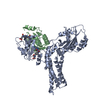
| Title | Crystal structure of the human dual specificity 1 catalytic domain (C258S) as a maltose binding protein fusion in complex with the designed AR protein mbp3_16 | ||||||
 Components Components |
| ||||||
 Keywords Keywords | HYDROLASE / DUSP / C258S / MBP / DARPin | ||||||
| Function / homology |  Function and homology information Function and homology informationnegative regulation of monocyte chemotaxis / peptidyl-serine dephosphorylation / endoderm formation / MAP kinase tyrosine/serine/threonine phosphatase activity / protein tyrosine/serine/threonine phosphatase activity / negative regulation of DNA biosynthetic process / peptidyl-threonine dephosphorylation / negative regulation of meiotic cell cycle / regulation of mitotic cell cycle spindle assembly checkpoint / negative regulation of p38MAPK cascade ...negative regulation of monocyte chemotaxis / peptidyl-serine dephosphorylation / endoderm formation / MAP kinase tyrosine/serine/threonine phosphatase activity / protein tyrosine/serine/threonine phosphatase activity / negative regulation of DNA biosynthetic process / peptidyl-threonine dephosphorylation / negative regulation of meiotic cell cycle / regulation of mitotic cell cycle spindle assembly checkpoint / negative regulation of p38MAPK cascade / RAF-independent MAPK1/3 activation / negative regulation of cell adhesion / cellular response to chemokine / mitogen-activated protein kinase binding / protein-serine/threonine phosphatase / response to testosterone / growth factor binding / detection of maltose stimulus / maltose transport complex / protein serine/threonine phosphatase activity / carbohydrate transport / response to light stimulus / response to retinoic acid / carbohydrate transmembrane transporter activity / maltose binding / response to cAMP / maltose transport / maltodextrin transmembrane transport / cellular response to hormone stimulus / ATP-binding cassette (ABC) transporter complex, substrate-binding subunit-containing / negative regulation of MAPK cascade / phosphoprotein phosphatase activity / protein-tyrosine-phosphatase / protein tyrosine phosphatase activity / ATP-binding cassette (ABC) transporter complex / response to glucocorticoid / cell chemotaxis / response to hydrogen peroxide / negative regulation of ERK1 and ERK2 cascade / response to calcium ion / Negative regulation of MAPK pathway / response to estradiol / outer membrane-bounded periplasmic space / periplasmic space / intracellular signal transduction / positive regulation of apoptotic process / negative regulation of cell population proliferation / DNA damage response / negative regulation of apoptotic process / signal transduction / membrane / nucleus / cytoplasm Similarity search - Function | ||||||
| Biological species |   Homo sapiens (human) Homo sapiens (human)synthetic construct (others) | ||||||
| Method |  X-RAY DIFFRACTION / MOLECULAR REPLACEMENT / Resolution: 2.226 Å X-RAY DIFFRACTION / MOLECULAR REPLACEMENT / Resolution: 2.226 Å | ||||||
 Authors Authors | Gumpena, R. / Waugh, D.S. / Lountos, G.T. | ||||||
 Citation Citation | Journal: Acta Crystallogr F Struct Biol Commun / Year: 2018 Title: MBP-binding DARPins facilitate the crystallization of an MBP fusion protein. Authors: Gumpena, R. / Lountos, G.T. / Waugh, D.S. | ||||||
| History |
|
- Structure visualization
| Structure viewer | Molecule: MolmilJmol/JSmol |
|---|
- Downloads & links
-Download
| PDBx/mmCIF format | 6d66.cif.gz | 151.2 KB | Display | PDBx/mmCIF format |
|---|---|---|---|---|
| PDB format | pdb6d66.ent.gz | 115.2 KB | Display | PDB format |
| PDBx/mmJSON format | 6d66.json.gz | Tree view | PDBx/mmJSON format | |
| Others |  Other downloads Other downloads |
-Validation report
| Arichive directory | https://data.pdbj.org/pub/pdb/validation_reports/d6/6d66ftp://data.pdbj.org/pub/pdb/validation_reports/d6/6d66 | HTTPS FTP |
|---|
-Related structure data
| Related structure data |  6d65SC  6d67C S: Starting model for refinement C: citing same article ( |
|---|---|
| Similar structure data |


-Links
 PDBj
PDBj


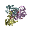
- Assembly
| Deposited unit | 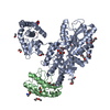
| ||||||||
|---|---|---|---|---|---|---|---|---|---|
| 1 |
| ||||||||
| Unit cell |
|
-Components
-Protein , 2 types, 2 molecules AB
| #1: Protein | Mass: 57103.613 Da / Num. of mol.: 1 Mutation: D82A, K83A, E172A, N173A, K239A, E362A, D363A, C258S,D82A, K83A, E172A, N173A, K239A, E362A, D363A, C258S Source method: isolated from a genetically manipulated source Source: (gene. exp.) Homo sapiens (human)Strain: K12 / Gene: malE, b4034, JW3994, DUSP1, CL100, MKP1, PTPN10, VH1 / Production host: References: UniProt: P0AEX9, UniProt: P28562, protein-serine/threonine phosphatase, protein-tyrosine-phosphatase |
|---|---|
| #2: Protein | Mass: 14787.509 Da / Num. of mol.: 1 Source method: isolated from a genetically manipulated source Source: (gene. exp.) synthetic construct (others) / Production host: |
-Non-polymers , 8 types, 331 molecules 


| #3: Chemical |  Mass: 106.120 Da / Num. of mol.: 3 / Source method: obtained synthetically / Formula: C4H10O3 Mass: 106.120 Da / Num. of mol.: 3 / Source method: obtained synthetically / Formula: C4H10O3#4: Chemical | ChemComp-PO4 / |  Mass: 94.971 Da / Num. of mol.: 1 / Source method: obtained synthetically / Formula: PO4 Mass: 94.971 Da / Num. of mol.: 1 / Source method: obtained synthetically / Formula: PO4#5: Chemical |  Type: peptide linking / Mass: 75.067 Da / Num. of mol.: 3 / Source method: obtained synthetically / Formula: C2H5NO2 Type: peptide linking / Mass: 75.067 Da / Num. of mol.: 3 / Source method: obtained synthetically / Formula: C2H5NO2#6: Chemical |  Mass: 150.173 Da / Num. of mol.: 3 / Source method: isolated from a natural source / Formula: C6H14O4 Mass: 150.173 Da / Num. of mol.: 3 / Source method: isolated from a natural source / Formula: C6H14O4#7: Chemical | ChemComp-EDO /  Mass: 62.068 Da / Num. of mol.: 12 / Source method: obtained synthetically / Formula: C2H6O2 Mass: 62.068 Da / Num. of mol.: 12 / Source method: obtained synthetically / Formula: C2H6O2#8: Chemical | ChemComp-PG4 / |  Mass: 194.226 Da / Num. of mol.: 1 / Source method: obtained synthetically / Formula: C8H18O5 / Comment: precipitant*YM Mass: 194.226 Da / Num. of mol.: 1 / Source method: obtained synthetically / Formula: C8H18O5 / Comment: precipitant*YM#9: Chemical | ChemComp-DAL / |  Type: D-peptide linking / Mass: 89.093 Da / Num. of mol.: 1 / Source method: obtained synthetically / Formula: C3H7NO2 Type: D-peptide linking / Mass: 89.093 Da / Num. of mol.: 1 / Source method: obtained synthetically / Formula: C3H7NO2#10: Water | ChemComp-HOH / | Mass: 18.015 Da / Num. of mol.: 307 / Source method: isolated from a natural source / Formula: H2O |
|---|
-Experimental details
-Experiment
| Experiment | Method: X-RAY DIFFRACTION / Number of used crystals: 1 |
|---|
- Sample preparation
| Crystal | Density Matthews: 2.96 Å3/Da / Density % sol: 58.38 % |
|---|---|
| Crystal grow | Temperature: 292 K / Method: vapor diffusion, hanging drop / pH: 8.5 Details: 0.2 M DL-glutamic acid 0.2 M DL-alanine 0.2 M -glycine 0.2 M-DL-lysine 0.2 M DL-serine 0.1 M Tris: Bicine 25% MPD 25% PEG1000 25% PEG3350 |
-Data collection
| Diffraction | Mean temperature: 100 K |
|---|---|
| Diffraction source | Source: ROTATING ANODE / Type: RIGAKU MICROMAX-007 HF / Wavelength: 1.54 Å |
| Detector | Type: MARRESEARCH / Detector: CCD / Date: May 11, 2016 |
| Radiation | Monochromator: Cu / Protocol: SINGLE WAVELENGTH / Monochromatic (M) / Laue (L): M / Scattering type: x-ray |
| Radiation wavelength | Wavelength: 1.54 Å / Relative weight: 1 |
| Reflection | Resolution: 2.22→50 Å / Num. obs: 43247 / % possible obs: 98.6 % / Redundancy: 10.7 % / Rmerge(I) obs: 0.05 / Net I/σ(I): 46.4 |
| Reflection shell | Resolution: 2.22→2.3 Å / Redundancy: 7.7 % / Rmerge(I) obs: 0.388 / Mean I/σ(I) obs: 3.6 / Num. unique obs: 3697 / % possible all: 86.5 |
- Processing
| Software |
| ||||||||||||||||||||||||||||||||||||||||||||||||||||||||||||||||||||||||||||||||||||||||||||||||||||||||||||||||
|---|---|---|---|---|---|---|---|---|---|---|---|---|---|---|---|---|---|---|---|---|---|---|---|---|---|---|---|---|---|---|---|---|---|---|---|---|---|---|---|---|---|---|---|---|---|---|---|---|---|---|---|---|---|---|---|---|---|---|---|---|---|---|---|---|---|---|---|---|---|---|---|---|---|---|---|---|---|---|---|---|---|---|---|---|---|---|---|---|---|---|---|---|---|---|---|---|---|---|---|---|---|---|---|---|---|---|---|---|---|---|---|---|---|
| Refinement | Method to determine structure: MOLECULAR REPLACEMENT Starting model: 6D65 Resolution: 2.226→36.444 Å / SU ML: 0.25 / Cross valid method: THROUGHOUT / σ(F): 1.35 / Phase error: 18.42
| ||||||||||||||||||||||||||||||||||||||||||||||||||||||||||||||||||||||||||||||||||||||||||||||||||||||||||||||||
| Solvent computation | Shrinkage radii: 0.9 Å / VDW probe radii: 1.11 Å | ||||||||||||||||||||||||||||||||||||||||||||||||||||||||||||||||||||||||||||||||||||||||||||||||||||||||||||||||
| Refinement step | Cycle: LAST / Resolution: 2.226→36.444 Å
| ||||||||||||||||||||||||||||||||||||||||||||||||||||||||||||||||||||||||||||||||||||||||||||||||||||||||||||||||
| Refine LS restraints |
| ||||||||||||||||||||||||||||||||||||||||||||||||||||||||||||||||||||||||||||||||||||||||||||||||||||||||||||||||
| LS refinement shell |
|
