 Movie
Movie Controller

[English] 日本語
 Yorodumi
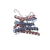
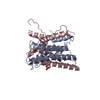
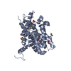
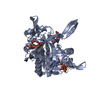
- PDB-6cwx: Crystal structure of human ribonuclease P/MRP proteins Rpp20/Rpp25 -
+ Open data

- Basic information
| Entry | Database: PDB / ID: 6cwx | ||||||||||||
|---|---|---|---|---|---|---|---|---|---|---|---|---|---|
| Title | Crystal structure of human ribonuclease P/MRP proteins Rpp20/Rpp25 | ||||||||||||
 Components Components |
| ||||||||||||
 Keywords Keywords | HYDROLASE / endonuclease / ribonuclease P / ribonuclease P complex / ribonuclease MRP / ribonuclease MRP complex / tRNA processing / rRNA processing / nucleic acid binding / RNA binding / protein binding / protein heterodimer / heterodimer / dimer / nucleus / nucleolus | ||||||||||||
| Function / homology |  Function and homology information Function and homology informationmultimeric ribonuclease P complex / nucleolar ribonuclease P complex / ribonuclease P RNA binding / ribonuclease MRP complex / tRNA processing in the nucleus / tRNA 5'-leader removal / tRNA processing / Major pathway of rRNA processing in the nucleolus and cytosol / centriolar satellite / rRNA processing ...multimeric ribonuclease P complex / nucleolar ribonuclease P complex / ribonuclease P RNA binding / ribonuclease MRP complex / tRNA processing in the nucleus / tRNA 5'-leader removal / tRNA processing / Major pathway of rRNA processing in the nucleolus and cytosol / centriolar satellite / rRNA processing / nucleolus / RNA binding / nucleoplasm / nucleus / cytoplasm Similarity search - Function | ||||||||||||
| Biological species |  Homo sapiens (human) Homo sapiens (human) | ||||||||||||
| Method |  X-RAY DIFFRACTION / SYNCHROTRON / SIRAS / Resolution: 2.25 Å X-RAY DIFFRACTION / SYNCHROTRON / SIRAS / Resolution: 2.25 Å | ||||||||||||
 Authors Authors | Chan, C.W. / Kiesel, B.R. / Mondragon, A. | ||||||||||||
| Funding support |  United States, 3items United States, 3items
| ||||||||||||
 Citation Citation | Journal: J. Mol. Biol. / Year: 2018 Title: Crystal Structure of Human Rpp20/Rpp25 Reveals Quaternary Level Adaptation of the Alba Scaffold as Structural Basis for Single-stranded RNA Binding. Authors: Chan, C.W. / Kiesel, B.R. / Mondragon, A. | ||||||||||||
| History |
|
- Structure visualization
| Structure viewer | Molecule: MolmilJmol/JSmol |
|---|
- Downloads & links
-Download
| PDBx/mmCIF format | 6cwx.cif.gz | 107.8 KB | Display | PDBx/mmCIF format |
|---|---|---|---|---|
| PDB format | pdb6cwx.ent.gz | 83 KB | Display | PDB format |
| PDBx/mmJSON format | 6cwx.json.gz | Tree view | PDBx/mmJSON format | |
| Others |  Other downloads Other downloads |
-Validation report
| Arichive directory | https://data.pdbj.org/pub/pdb/validation_reports/cw/6cwxftp://data.pdbj.org/pub/pdb/validation_reports/cw/6cwx | HTTPS FTP |
|---|
-Related structure data
| Similar structure data |
|---|


-Links
 PDBj
PDBj


- Assembly
| Deposited unit | 
| ||||||||
|---|---|---|---|---|---|---|---|---|---|
| 1 |
| ||||||||
| 2 | x 24
| ||||||||
| Unit cell |
|
-Components
| #1: Protein | Mass: 16020.300 Da / Num. of mol.: 1 Source method: isolated from a genetically manipulated source Source: (gene. exp.) Homo sapiens (human) / Gene: POP7, RPP20 / Plasmid: pHTT7K / Production host:  | ||||||
|---|---|---|---|---|---|---|---|
| #2: Protein | Mass: 20930.723 Da / Num. of mol.: 1 Source method: isolated from a genetically manipulated source Source: (gene. exp.) Homo sapiens (human) / Gene: RPP25 / Plasmid: pHTT7K / Production host: | ||||||
| #3: Chemical |   Mass: 46.025 Da / Num. of mol.: 2 / Source method: obtained synthetically / Formula: CH2O2 Mass: 46.025 Da / Num. of mol.: 2 / Source method: obtained synthetically / Formula: CH2O2#4: Chemical |   Mass: 96.063 Da / Num. of mol.: 2 / Source method: obtained synthetically / Formula: SO4 Mass: 96.063 Da / Num. of mol.: 2 / Source method: obtained synthetically / Formula: SO4#5: Water | ChemComp-HOH / |  Mass: 18.015 Da / Num. of mol.: 100 / Source method: isolated from a natural source / Formula: H2O Mass: 18.015 Da / Num. of mol.: 100 / Source method: isolated from a natural source / Formula: H2OHas protein modification | Y | |
-Experimental details
-Experiment
| Experiment | Method: X-RAY DIFFRACTION / Number of used crystals: 1 |
|---|
- Sample preparation
| Crystal | Density Matthews: 3.42 Å3/Da / Density % sol: 63.93 % |
|---|---|
| Crystal grow | Temperature: 291.15 K / Method: vapor diffusion, hanging drop / pH: 7.5 Details: 100 mM potassium formate, pH 7.5, 300 mM magnesium sulfate, 4.5% PEG 300, 4.5% PEG 400, 4.5% PEG 1000, 4.5% PEG 4000, and 4.5% PEG 8000 PH range: 7.0 - 8.0 |
-Data collection
| Diffraction | Mean temperature: 100 K |
|---|---|
| Diffraction source | Source: SYNCHROTRON / Site: APS / Beamline: 21-ID-F / Wavelength: 0.97872 Å |
| Detector | Type: MARMOSAIC 225 mm CCD / Detector: CCD / Date: Jun 29, 2015 |
| Radiation | Protocol: SINGLE WAVELENGTH / Monochromatic (M) / Laue (L): M / Scattering type: x-ray |
| Radiation wavelength | Wavelength: 0.97872 Å / Relative weight: 1 |
| Reflection | Resolution: 2.25→48.69 Å / Num. obs: 24742 / % possible obs: 100 % / Redundancy: 9.7 % / CC1/2: 0.999 / Rmerge(I) obs: 0.083 / Rpim(I) all: 0.028 / Rrim(I) all: 0.087 / Net I/σ(I): 16.4 |
| Reflection shell | Resolution: 2.25→2.32 Å / Redundancy: 9.8 % / Rmerge(I) obs: 0.495 / Mean I/σ(I) obs: 4.7 / Num. unique obs: 2265 / CC1/2: 0.924 / Rpim(I) all: 0.165 / Rrim(I) all: 0.523 / % possible all: 100 |
-Phasing
| Phasing | Method: SIRAS |
|---|
- Processing
| Software |
| |||||||||||||||||||||||||||||||||||||||||||||||||||||||||||||||||||||||||||
|---|---|---|---|---|---|---|---|---|---|---|---|---|---|---|---|---|---|---|---|---|---|---|---|---|---|---|---|---|---|---|---|---|---|---|---|---|---|---|---|---|---|---|---|---|---|---|---|---|---|---|---|---|---|---|---|---|---|---|---|---|---|---|---|---|---|---|---|---|---|---|---|---|---|---|---|---|
| Refinement | Method to determine structure: SIRAS / Resolution: 2.25→48.69 Å / Cor.coef. Fo:Fc: 0.942 / Cor.coef. Fo:Fc free: 0.921 / SU B: 8.799 / SU ML: 0.115 / Cross valid method: THROUGHOUT / σ(F): 0 / ESU R: 0.166 / ESU R Free: 0.15 / Stereochemistry target values: MAXIMUM LIKELIHOOD Details: HYDROGENS HAVE BEEN ADDED IN THE RIDING POSITIONS U VALUES : WITH TLS ADDED
| |||||||||||||||||||||||||||||||||||||||||||||||||||||||||||||||||||||||||||
| Solvent computation | Ion probe radii: 0.8 Å / Shrinkage radii: 0.8 Å / VDW probe radii: 1.2 Å / Solvent model: MASK | |||||||||||||||||||||||||||||||||||||||||||||||||||||||||||||||||||||||||||
| Displacement parameters | Biso max: 135.26 Å2 / Biso mean: 54.096 Å2 / Biso min: 15.31 Å2
| |||||||||||||||||||||||||||||||||||||||||||||||||||||||||||||||||||||||||||
| Refinement step | Cycle: final / Resolution: 2.25→48.69 Å
| |||||||||||||||||||||||||||||||||||||||||||||||||||||||||||||||||||||||||||
| Refine LS restraints |
| |||||||||||||||||||||||||||||||||||||||||||||||||||||||||||||||||||||||||||
| LS refinement shell | Resolution: 2.251→2.309 Å / Rfactor Rfree error: 0 / Total num. of bins used: 20
| |||||||||||||||||||||||||||||||||||||||||||||||||||||||||||||||||||||||||||
| Refinement TLS params. | Method: refined / Refine-ID: X-RAY DIFFRACTION
| |||||||||||||||||||||||||||||||||||||||||||||||||||||||||||||||||||||||||||
| Refinement TLS group |
|
