 Movie
Movie Controller

[English] 日本語
 Yorodumi
- PDB-6cia: Crystal structure of aldo-keto reductase from Klebsiella pneumoni... -
+ Open data

- Basic information
| Entry | Database: PDB / ID: 6cia | ||||||
|---|---|---|---|---|---|---|---|
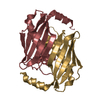
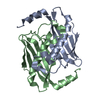
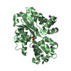
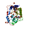
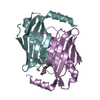
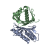
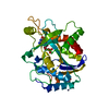
| Title | Crystal structure of aldo-keto reductase from Klebsiella pneumoniae in complex with NADPH. | ||||||
 Components Components | Aldo/keto reductase | ||||||
 Keywords Keywords | OXIDOREDUCTASE / Structural Genomics / PSI-Biology / Center for Structural Genomics of Infectious Diseases / CSGID | ||||||
| Function / homology |  Function and homology information Function and homology informationOxidoreductases; Acting on the CH-OH group of donors; With NAD+ or NADP+ as acceptor / oxidoreductase activity / nucleotide binding Similarity search - Function | ||||||
| Biological species |  Klebsiella pneumoniae (bacteria) Klebsiella pneumoniae (bacteria) | ||||||
| Method |  X-RAY DIFFRACTION / SYNCHROTRON / MOLECULAR REPLACEMENT / Resolution: 2.3 Å X-RAY DIFFRACTION / SYNCHROTRON / MOLECULAR REPLACEMENT / Resolution: 2.3 Å | ||||||
 Authors Authors | Lipowska, J. / Leung, E.S. / Shabalin, I.G. / Grabowski, M. / Almo, S.C. / Satchell, K.J. / Joachimiak, A. / Lewinski, K. / Minor, W. / Center for Structural Genomics of Infectious Diseases (CSGID) | ||||||
| Funding support |  United States, 1items United States, 1items
| ||||||
 Citation Citation | Journal: to be published Title: Crystal structure of of aldo-keto reductase from Klebsiella pneumoniae in complex with NADPH. Authors: Lipowska, J. / Leung, E.S. / Shabalin, I.G. / Grabowski, M. / Almo, S.C. / Satchell, K.J. / Joachimiak, A. / Lewinski, K. / Minor, W. | ||||||
| History |
|
- Structure visualization
| Structure viewer | Molecule: MolmilJmol/JSmol |
|---|
- Downloads & links
-Download
| PDBx/mmCIF format | 6cia.cif.gz | 134.6 KB | Display | PDBx/mmCIF format |
|---|---|---|---|---|
| PDB format | pdb6cia.ent.gz | 103.6 KB | Display | PDB format |
| PDBx/mmJSON format | 6cia.json.gz | Tree view | PDBx/mmJSON format | |
| Others |  Other downloads Other downloads |
-Validation report
| Arichive directory | https://data.pdbj.org/pub/pdb/validation_reports/ci/6ciaftp://data.pdbj.org/pub/pdb/validation_reports/ci/6cia | HTTPS FTP |
|---|
-Related structure data
| Related structure data | 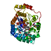 4wghS S: Starting model for refinement |
|---|---|
| Similar structure data | |
| Other databases |


-Links
 PDBj
PDBj


- Assembly
| Deposited unit | 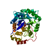
| ||||||||
|---|---|---|---|---|---|---|---|---|---|
| 1 |
| ||||||||
| Unit cell |
|
-Components
| #1: Protein | Mass: 31768.480 Da / Num. of mol.: 1 Source method: isolated from a genetically manipulated source Source: (gene. exp.) Klebsiella pneumoniae (bacteria)Gene: yhdN_4, yhdN, B1727_06910, B4U21_09420, BN49_2327, CAK82_20800, CQB04_18905, CR230_19100, CTI52_15935, CTI54_15930, PMK1_03573, SAMEA3531778_00013 Production host: References: UniProt: W9BFN4, UniProt: A6T7Q7*PLUS, Oxidoreductases; Acting on the CH-OH group of donors; With NAD+ or NADP+ as acceptor |
|---|---|
| #2: Chemical | ChemComp-NDP /   Mass: 745.421 Da / Num. of mol.: 1 / Source method: obtained synthetically / Formula: C21H30N7O17P3 Mass: 745.421 Da / Num. of mol.: 1 / Source method: obtained synthetically / Formula: C21H30N7O17P3 |
| #3: Water | ChemComp-HOH /  Mass: 18.015 Da / Num. of mol.: 150 / Source method: isolated from a natural source / Formula: H2O Mass: 18.015 Da / Num. of mol.: 150 / Source method: isolated from a natural source / Formula: H2O |
-Experimental details
-Experiment
| Experiment | Method: X-RAY DIFFRACTION / Number of used crystals: 1 |
|---|
- Sample preparation
| Crystal | Density Matthews: 2.31 Å3/Da / Density % sol: 46.85 % |
|---|---|
| Crystal grow | Temperature: 289.15 K / Method: vapor diffusion, sitting drop / pH: 8.5 Details: 0.2 ul of 11 mg/ml protein in 20 mM HEPES pH 7.5, 150 mM NaCl, 10% Glycerol, 0.1% Sodium Azide and 0.5 mM TCEP were mixed with 0.2 ul of the Top 96 #90 (0.2 uL 0.1 M Tris: HCl, pH 8.5, 25 % ...Details: 0.2 ul of 11 mg/ml protein in 20 mM HEPES pH 7.5, 150 mM NaCl, 10% Glycerol, 0.1% Sodium Azide and 0.5 mM TCEP were mixed with 0.2 ul of the Top 96 #90 (0.2 uL 0.1 M Tris: HCl, pH 8.5, 25 % (w/v) PEG 3350) and 0.1 uL 30%v/v Ethanol (Additive Screen #82) and equilibrated against 1.5 M NaCl solution in 96 Well 3 drop Crystallization Plate (Swissci) |
-Data collection
| Diffraction | Mean temperature: 100 K |
|---|---|
| Diffraction source | Source: SYNCHROTRON / Site: APS / Beamline: 21-ID-G / Wavelength: 0.97856 Å |
| Detector | Type: MARMOSAIC 300 mm CCD / Detector: CCD / Date: Apr 8, 2017 / Details: mirrors |
| Radiation | Protocol: SINGLE WAVELENGTH / Monochromatic (M) / Laue (L): M / Scattering type: x-ray |
| Radiation wavelength | Wavelength: 0.97856 Å / Relative weight: 1 |
| Reflection | Resolution: 2.3→50 Å / Num. obs: 13360 / % possible obs: 100 % / Observed criterion σ(I): -3 / Redundancy: 8.3 % / Biso Wilson estimate: 33.7 Å2 / CC1/2: 1 / Rmerge(I) obs: 0.083 / Rpim(I) all: 0.03 / Rrim(I) all: 0.088 / Rsym value: 0.083 / Net I/av σ(I): 30.9 / Net I/σ(I): 30.9 |
| Reflection shell | Resolution: 2.3→2.34 Å / Redundancy: 7 % / Rmerge(I) obs: 1.092 / Mean I/σ(I) obs: 1.9 / Num. unique obs: 649 / CC1/2: 0.838 / Rpim(I) all: 0.438 / Rrim(I) all: 1.178 / Rsym value: 1.092 / % possible all: 100 |
- Processing
| Software |
| ||||||||||||||||||||||||||||||||||||||||||||||||||||||||||||||||||||||||||||||||||||||||||||||||||||||||||||||||||||||||||||||||||||||||||||||||||||||||||||||||||||||||||||||||||||||
|---|---|---|---|---|---|---|---|---|---|---|---|---|---|---|---|---|---|---|---|---|---|---|---|---|---|---|---|---|---|---|---|---|---|---|---|---|---|---|---|---|---|---|---|---|---|---|---|---|---|---|---|---|---|---|---|---|---|---|---|---|---|---|---|---|---|---|---|---|---|---|---|---|---|---|---|---|---|---|---|---|---|---|---|---|---|---|---|---|---|---|---|---|---|---|---|---|---|---|---|---|---|---|---|---|---|---|---|---|---|---|---|---|---|---|---|---|---|---|---|---|---|---|---|---|---|---|---|---|---|---|---|---|---|---|---|---|---|---|---|---|---|---|---|---|---|---|---|---|---|---|---|---|---|---|---|---|---|---|---|---|---|---|---|---|---|---|---|---|---|---|---|---|---|---|---|---|---|---|---|---|---|---|---|
| Refinement | Method to determine structure: MOLECULAR REPLACEMENT Starting model: 4WGH Resolution: 2.3→50 Å / Cor.coef. Fo:Fc: 0.959 / Cor.coef. Fo:Fc free: 0.924 / SU B: 15.302 / SU ML: 0.201 / Cross valid method: THROUGHOUT / ESU R: 0.49 / ESU R Free: 0.257 / Details: HYDROGENS HAVE BEEN ADDED IN THE RIDING POSITIONS
| ||||||||||||||||||||||||||||||||||||||||||||||||||||||||||||||||||||||||||||||||||||||||||||||||||||||||||||||||||||||||||||||||||||||||||||||||||||||||||||||||||||||||||||||||||||||
| Solvent computation | Ion probe radii: 0.8 Å / Shrinkage radii: 0.8 Å / VDW probe radii: 1.2 Å | ||||||||||||||||||||||||||||||||||||||||||||||||||||||||||||||||||||||||||||||||||||||||||||||||||||||||||||||||||||||||||||||||||||||||||||||||||||||||||||||||||||||||||||||||||||||
| Displacement parameters | Biso mean: 43.872 Å2
| ||||||||||||||||||||||||||||||||||||||||||||||||||||||||||||||||||||||||||||||||||||||||||||||||||||||||||||||||||||||||||||||||||||||||||||||||||||||||||||||||||||||||||||||||||||||
| Refinement step | Cycle: 1 / Resolution: 2.3→50 Å
| ||||||||||||||||||||||||||||||||||||||||||||||||||||||||||||||||||||||||||||||||||||||||||||||||||||||||||||||||||||||||||||||||||||||||||||||||||||||||||||||||||||||||||||||||||||||
| Refine LS restraints |
|