 Movie
Movie Controller

[English] 日本語
 Yorodumi
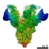
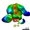
- PDB-6chb: Crystal structure of a natively-glycosylated BG505 SOSIP.664 HIV-... -
+ Open data

- Basic information
| Entry | Database: PDB / ID: 6chb | ||||||||||||
|---|---|---|---|---|---|---|---|---|---|---|---|---|---|
| Title | Crystal structure of a natively-glycosylated BG505 SOSIP.664 HIV-1 Envelope Trimer in complex with the broadly-neutralizing antibodies BG18 and IOMA | ||||||||||||
 Components Components |
| ||||||||||||
 Keywords Keywords | IMMUNE SYSTEM / Env glycoprotein / broadly neutralizing antibodies | ||||||||||||
| Function / homology |  Function and homology information Function and homology informationsymbiont-mediated perturbation of host defense response / positive regulation of plasma membrane raft polarization / positive regulation of receptor clustering / host cell endosome membrane / clathrin-dependent endocytosis of virus by host cell / viral protein processing / fusion of virus membrane with host plasma membrane / fusion of virus membrane with host endosome membrane / viral envelope / virion attachment to host cell ...symbiont-mediated perturbation of host defense response / positive regulation of plasma membrane raft polarization / positive regulation of receptor clustering / host cell endosome membrane / clathrin-dependent endocytosis of virus by host cell / viral protein processing / fusion of virus membrane with host plasma membrane / fusion of virus membrane with host endosome membrane / viral envelope / virion attachment to host cell / host cell plasma membrane / virion membrane / structural molecule activity / membrane / identical protein binding Similarity search - Function | ||||||||||||
| Biological species |   Human immunodeficiency virus 1 Human immunodeficiency virus 1 Homo sapiens (human) Homo sapiens (human) | ||||||||||||
| Method |  X-RAY DIFFRACTION / SYNCHROTRON / MOLECULAR REPLACEMENT / Resolution: 6.801 Å X-RAY DIFFRACTION / SYNCHROTRON / MOLECULAR REPLACEMENT / Resolution: 6.801 Å | ||||||||||||
 Authors Authors | Barnes, C.O. / Bjorkman, P.J. | ||||||||||||
| Funding support |  United States, 3items United States, 3items
| ||||||||||||
 Citation Citation | Journal: Nat Commun / Year: 2018 Title: Structural characterization of a highly-potent V3-glycan broadly neutralizing antibody bound to natively-glycosylated HIV-1 envelope. Authors: Barnes, C.O. / Gristick, H.B. / Freund, N.T. / Escolano, A. / Lyubimov, A.Y. / Hartweger, H. / West, A.P. / Cohen, A.E. / Nussenzweig, M.C. / Bjorkman, P.J. | ||||||||||||
| History |
|
- Structure visualization
| Structure viewer | Molecule: MolmilJmol/JSmol |
|---|
- Downloads & links
-Download
| PDBx/mmCIF format | 6chb.cif.gz | 745.1 KB | Display | PDBx/mmCIF format |
|---|---|---|---|---|
| PDB format | pdb6chb.ent.gz | 596.5 KB | Display | PDB format |
| PDBx/mmJSON format | 6chb.json.gz | Tree view | PDBx/mmJSON format | |
| Others |  Other downloads Other downloads |
-Validation report
| Arichive directory | https://data.pdbj.org/pub/pdb/validation_reports/ch/6chbftp://data.pdbj.org/pub/pdb/validation_reports/ch/6chb | HTTPS FTP |
|---|
-Related structure data
| Related structure data | 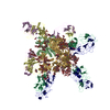 6ch7C  6ch8C  6ch9C  5t3zS 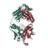 5ud9S S: Starting model for refinement C: citing same article ( |
|---|---|
| Similar structure data |


-Links
 PDBj
PDBj


- Assembly
| Deposited unit | 
| ||||||||
|---|---|---|---|---|---|---|---|---|---|
| 1 |
| ||||||||
| Unit cell |
|
-Components
-Envelope glycoprotein ... , 2 types, 6 molecules BACGFH
| #1: Protein | Mass: 17162.525 Da / Num. of mol.: 3 Source method: isolated from a genetically manipulated source Source: (gene. exp.) Human immunodeficiency virus 1 / Gene: env / Plasmid: pAM/C / Production host:   Cricetulus griseus (Chinese hamster) / Strain (production host): K1 / References: UniProt: Q2N0S7, UniProt: Q2N0S5*PLUS Cricetulus griseus (Chinese hamster) / Strain (production host): K1 / References: UniProt: Q2N0S7, UniProt: Q2N0S5*PLUS#2: Protein | Mass: 53693.789 Da / Num. of mol.: 3 Source method: isolated from a genetically manipulated source Source: (gene. exp.) Human immunodeficiency virus 1 / Gene: env / Plasmid: pAM/C / Production host: Cricetulus griseus (Chinese hamster) / Strain (production host): K1 / References: UniProt: Q2N0S6 |
|---|
-Protein , 1 types, 3 molecules JIQ
| #3: Protein | Mass: 26158.375 Da / Num. of mol.: 3 Source method: isolated from a genetically manipulated source Source: (gene. exp.) Homo sapiens (human) / Production host: Homo sapiens (human) |
|---|
-Antibody , 3 types, 9 molecules KLRDMOENP
| #4: Antibody | Mass: 23021.447 Da / Num. of mol.: 3 Source method: isolated from a genetically manipulated source Source: (gene. exp.) Homo sapiens (human) / Production host: Homo sapiens (human)#5: Antibody | Mass: 25176.416 Da / Num. of mol.: 3 Source method: isolated from a genetically manipulated source Source: (gene. exp.) Homo sapiens (human) / Plasmid: pTT5 / Cell line (production host): HEK293-6E / Production host: Homo sapiens (human)#6: Antibody | Mass: 22591.975 Da / Num. of mol.: 3 Source method: isolated from a genetically manipulated source Source: (gene. exp.) Homo sapiens (human) / Plasmid: pTT5 / Cell line (production host): HEK293-6E / Production host: Homo sapiens (human) |
|---|
-Details
| Has protein modification | Y |
|---|
-Experimental details
-Experiment
| Experiment | Method: X-RAY DIFFRACTION / Number of used crystals: 1 |
|---|
- Sample preparation
| Crystal | Density Matthews: 3.55 Å3/Da / Density % sol: 65.39 % / Mosaicity: 0.22 ° |
|---|---|
| Crystal grow | Temperature: 298 K / Method: vapor diffusion, hanging drop / pH: 8 / Details: 5% Tacismate pH 8.0, 15% PEG 3350 |
-Data collection
| Diffraction | Mean temperature: 100 K | ||||||||||||||||||||||||
|---|---|---|---|---|---|---|---|---|---|---|---|---|---|---|---|---|---|---|---|---|---|---|---|---|---|
| Diffraction source | Source: SYNCHROTRON / Site: SSRL / Beamline: BL12-2 / Wavelength: 1 Å | ||||||||||||||||||||||||
| Detector | Type: DECTRIS PILATUS 6M / Detector: PIXEL / Date: Apr 26, 2017 | ||||||||||||||||||||||||
| Radiation | Protocol: SINGLE WAVELENGTH / Monochromatic (M) / Laue (L): M / Scattering type: x-ray | ||||||||||||||||||||||||
| Radiation wavelength | Wavelength: 1 Å / Relative weight: 1 | ||||||||||||||||||||||||
| Reflection | Resolution: 6.66→39.7 Å / Num. obs: 13894 / % possible obs: 98.7 % / Redundancy: 12.4 % / Biso Wilson estimate: 368.33 Å2 / CC1/2: 0.988 / Rmerge(I) obs: 0.333 / Rpim(I) all: 0.097 / Rrim(I) all: 0.347 / Net I/σ(I): 6.4 | ||||||||||||||||||||||||
| Reflection shell | Diffraction-ID: 1
|
- Processing
| Software |
| |||||||||||||||||||||||||||||||||||||||||||||||||||||||||||||||
|---|---|---|---|---|---|---|---|---|---|---|---|---|---|---|---|---|---|---|---|---|---|---|---|---|---|---|---|---|---|---|---|---|---|---|---|---|---|---|---|---|---|---|---|---|---|---|---|---|---|---|---|---|---|---|---|---|---|---|---|---|---|---|---|---|
| Refinement | Method to determine structure: MOLECULAR REPLACEMENT Starting model: 5T3Z, 5UD9 Resolution: 6.801→39.631 Å / SU ML: 1.68 / Cross valid method: THROUGHOUT / σ(F): 0.23 / Phase error: 51.74 / Stereochemistry target values: ML
| |||||||||||||||||||||||||||||||||||||||||||||||||||||||||||||||
| Solvent computation | Shrinkage radii: 0.9 Å / VDW probe radii: 1.11 Å / Solvent model: FLAT BULK SOLVENT MODEL | |||||||||||||||||||||||||||||||||||||||||||||||||||||||||||||||
| Refinement step | Cycle: LAST / Resolution: 6.801→39.631 Å
| |||||||||||||||||||||||||||||||||||||||||||||||||||||||||||||||
| Refine LS restraints |
| |||||||||||||||||||||||||||||||||||||||||||||||||||||||||||||||
| LS refinement shell |
|
