 Movie
Movie Controller


+ Open data

- Basic information
| Entry | Database: PDB / ID: 6cfd | ||||||
|---|---|---|---|---|---|---|---|
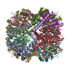
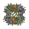
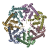
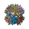
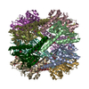
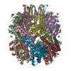
| Title | ADEP4 bound to E. faecium ClpP | ||||||
 Components Components | ATP-dependent Clp protease proteolytic subunit | ||||||
 Keywords Keywords | HYDROLASE/ANTIBIOTIC / ClpP / ADEP4 / HYDROLASE-ANTIBIOTIC complex | ||||||
| Function / homology |  Function and homology information Function and homology informationendopeptidase Clp / endopeptidase Clp complex / ATP-dependent peptidase activity / protein quality control for misfolded or incompletely synthesized proteins / ATPase binding / serine-type endopeptidase activity / cytoplasm Similarity search - Function | ||||||
| Biological species |  Enterococcus faecium (bacteria) Enterococcus faecium (bacteria) | ||||||
| Method |  X-RAY DIFFRACTION / SYNCHROTRON / MOLECULAR REPLACEMENT / Resolution: 2.57 Å X-RAY DIFFRACTION / SYNCHROTRON / MOLECULAR REPLACEMENT / Resolution: 2.57 Å | ||||||
 Authors Authors | Lee, R.E. / Griffith, E.C. | ||||||
| Funding support |  United States, 1items United States, 1items
| ||||||
 Citation Citation | Journal: Antimicrob. Agents Chemother. / Year: 2018 Title: In VivoandIn VitroEffects of a ClpP-Activating Antibiotic against Vancomycin-Resistant Enterococci. Authors: Brown Gandt, A. / Griffith, E.C. / Lister, I.M. / Billings, L.L. / Han, A. / Tangallapally, R. / Zhao, Y. / Singh, A.P. / Lee, R.E. / LaFleur, M.D. #1: Journal: J. Biol. Chem. / Year: 2011Title: Structural switching of Staphylococcus aureus Clp protease: a key to understanding protease dynamics. Authors: Zhang, J. / Ye, F. / Lan, L. / Jiang, H. / Luo, C. / Yang, C.G. #2: Journal: Chem. Biol. / Year: 2010Title: Acyldepsipeptide antibiotics induce the formation of a structured axial channel in ClpP: A model for the ClpX/ClpA-bound state of ClpP. Authors: Li, D.H. / Chung, Y.S. / Gloyd, M. / Joseph, E. / Ghirlando, R. / Wright, G.D. / Cheng, Y.Q. / Maurizi, M.R. / Guarne, A. / Ortega, J. #3: Journal: J. Biol. Chem. / Year: 2012Title: Insights into structural network responsible for oligomerization and activity of bacterial virulence regulator caseinolytic protease P (ClpP) protein. Authors: Gersch, M. / List, A. / Groll, M. / Sieber, S.A. | ||||||
| History |
|
- Structure visualization
| Structure viewer | Molecule: MolmilJmol/JSmol |
|---|
- Downloads & links
-Download
| PDBx/mmCIF format | 6cfd.cif.gz | 501.3 KB | Display | PDBx/mmCIF format |
|---|---|---|---|---|
| PDB format | pdb6cfd.ent.gz | 413.3 KB | Display | PDB format |
| PDBx/mmJSON format | 6cfd.json.gz | Tree view | PDBx/mmJSON format | |
| Others |  Other downloads Other downloads |
-Validation report
| Summary document | 6cfd_validation.pdf.gz | 3.7 MB | Display | wwPDB validaton report |
|---|---|---|---|---|
| Full document | 6cfd_full_validation.pdf.gz | 3.8 MB | Display | |
| Data in XML | 6cfd_validation.xml.gz | 99.7 KB | Display | |
| Data in CIF | 6cfd_validation.cif.gz | 123 KB | Display | |
| Arichive directory | https://data.pdbj.org/pub/pdb/validation_reports/cf/6cfdftp://data.pdbj.org/pub/pdb/validation_reports/cf/6cfd | HTTPS FTP |
-Related structure data
| Related structure data |  3staS S: Starting model for refinement |
|---|---|
| Similar structure data |


-Links
 PDBj
PDBj- Assembly
| Deposited unit | 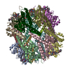
| ||||||||
|---|---|---|---|---|---|---|---|---|---|
| 1 |
| ||||||||
| Unit cell |
|
-Components
| #1: Protein | Mass: 23297.496 Da / Num. of mol.: 14 Source method: isolated from a genetically manipulated source Source: (gene. exp.) Enterococcus faecium (bacteria)Gene: clpP, A5804_002055, A5809_001747, A5810_000386, A5814_002098, A5836_000160, A5841_002116, A5860_002409, AS238_05290, AWT83_05460, B1P95_10385, B4W81_08585, BK413_10305, BK695_10280, BK696_ ...Gene: clpP, A5804_002055, A5809_001747, A5810_000386, A5814_002098, A5836_000160, A5841_002116, A5860_002409, AS238_05290, AWT83_05460, B1P95_10385, B4W81_08585, BK413_10305, BK695_10280, BK696_10275, BTA10_07855, BTA13_11970, BU181_02235, BU182_15300, BU184_14080, BU185_04695, BU186_06175, BU187_13865, BU188_14880, BU189_03420, BU190_07525, BU191_15420, BU192_05390, BU194_04805, CDL00_11615, CKY17_04320, CKY19_04880, CQR37_09710, CQR38_04150, CQR40_12365, CQR41_03640, CQR43_09890, CRM75_14685, CRN03_11890, CS913_12780, CS915_12510, CS916_12510, DTPHA_601708, EFM1CSP_03625, HMPREF3199_00581 Production host: References: UniProt: A0A133CH35, UniProt: Q3XX76*PLUS, endopeptidase Clp #2: Chemical | ChemComp-MPD / (   Mass: 118.174 Da / Num. of mol.: 14 / Source method: obtained synthetically / Formula: C6H14O2 / Comment: precipitant*YM Mass: 118.174 Da / Num. of mol.: 14 / Source method: obtained synthetically / Formula: C6H14O2 / Comment: precipitant*YM#3: Chemical | ChemComp-EZA /   Mass: 770.862 Da / Num. of mol.: 11 / Source method: obtained synthetically / Formula: C39H52F2N6O8 / Feature type: SUBJECT OF INVESTIGATION Mass: 770.862 Da / Num. of mol.: 11 / Source method: obtained synthetically / Formula: C39H52F2N6O8 / Feature type: SUBJECT OF INVESTIGATION#4: Water | ChemComp-HOH / |  Mass: 18.015 Da / Num. of mol.: 139 / Source method: isolated from a natural source / Formula: H2O Mass: 18.015 Da / Num. of mol.: 139 / Source method: isolated from a natural source / Formula: H2O |
|---|
-Experimental details
-Experiment
| Experiment | Method: X-RAY DIFFRACTION / Number of used crystals: 1 |
|---|
- Sample preparation
| Crystal | Density Matthews: 2.87 Å3/Da / Density % sol: 57.11 % / Description: A 0.3 mM cube forms within 72 hours. |
|---|---|
| Crystal grow | Temperature: 291.15 K / Method: vapor diffusion, hanging drop / pH: 4.5 Details: 0.1 M sodium acetate, pH 4.5, 18-35% MPD, 0.02 M calcium chloride PH range: 4.5-4.5 |
-Data collection
| Diffraction | Mean temperature: 100 K | |||||||||||||||||||||||||||||||||||||||||||||||||||||||||||||||||||||||||||||||||||||||||||||||||||||||||||||||||||||||||||||||||||||||||||||||||||||||||||||||||||||||||||||||||||||||||||||
|---|---|---|---|---|---|---|---|---|---|---|---|---|---|---|---|---|---|---|---|---|---|---|---|---|---|---|---|---|---|---|---|---|---|---|---|---|---|---|---|---|---|---|---|---|---|---|---|---|---|---|---|---|---|---|---|---|---|---|---|---|---|---|---|---|---|---|---|---|---|---|---|---|---|---|---|---|---|---|---|---|---|---|---|---|---|---|---|---|---|---|---|---|---|---|---|---|---|---|---|---|---|---|---|---|---|---|---|---|---|---|---|---|---|---|---|---|---|---|---|---|---|---|---|---|---|---|---|---|---|---|---|---|---|---|---|---|---|---|---|---|---|---|---|---|---|---|---|---|---|---|---|---|---|---|---|---|---|---|---|---|---|---|---|---|---|---|---|---|---|---|---|---|---|---|---|---|---|---|---|---|---|---|---|---|---|---|---|---|---|---|
| Diffraction source | Source: SYNCHROTRON / Site: APS / Beamline: 22-ID / Wavelength: 1 Å | |||||||||||||||||||||||||||||||||||||||||||||||||||||||||||||||||||||||||||||||||||||||||||||||||||||||||||||||||||||||||||||||||||||||||||||||||||||||||||||||||||||||||||||||||||||||||||||
| Detector | Type: MARMOSAIC 300 mm CCD / Detector: CCD / Date: Jun 5, 2017 | |||||||||||||||||||||||||||||||||||||||||||||||||||||||||||||||||||||||||||||||||||||||||||||||||||||||||||||||||||||||||||||||||||||||||||||||||||||||||||||||||||||||||||||||||||||||||||||
| Radiation | Monochromator: double crystal Si(111) / Protocol: SINGLE WAVELENGTH / Monochromatic (M) / Laue (L): M / Scattering type: x-ray | |||||||||||||||||||||||||||||||||||||||||||||||||||||||||||||||||||||||||||||||||||||||||||||||||||||||||||||||||||||||||||||||||||||||||||||||||||||||||||||||||||||||||||||||||||||||||||||
| Radiation wavelength | Wavelength: 1 Å / Relative weight: 1 | |||||||||||||||||||||||||||||||||||||||||||||||||||||||||||||||||||||||||||||||||||||||||||||||||||||||||||||||||||||||||||||||||||||||||||||||||||||||||||||||||||||||||||||||||||||||||||||
| Reflection | Resolution: 2.6→50 Å / Num. obs: 114674 / % possible obs: 100 % / Redundancy: 6 % / Rmerge(I) obs: 0.163 / Rpim(I) all: 0.069 / Rrim(I) all: 0.178 / Χ2: 0.89 / Net I/σ(I): 7.4 / Num. measured all: 688990 | |||||||||||||||||||||||||||||||||||||||||||||||||||||||||||||||||||||||||||||||||||||||||||||||||||||||||||||||||||||||||||||||||||||||||||||||||||||||||||||||||||||||||||||||||||||||||||||
| Reflection shell | Diffraction-ID: 1
|
- Processing
| Software |
| ||||||||||||||||||||||||||||||||||||||||||||||||||||||||||||
|---|---|---|---|---|---|---|---|---|---|---|---|---|---|---|---|---|---|---|---|---|---|---|---|---|---|---|---|---|---|---|---|---|---|---|---|---|---|---|---|---|---|---|---|---|---|---|---|---|---|---|---|---|---|---|---|---|---|---|---|---|---|
| Refinement | Method to determine structure: MOLECULAR REPLACEMENT Starting model: PDB entry 3STA Resolution: 2.57→101.08 Å / Cor.coef. Fo:Fc: 0.959 / Cor.coef. Fo:Fc free: 0.944 / WRfactor Rfree: 0.2212 / WRfactor Rwork: 0.1945 / FOM work R set: 0.8361 / SU B: 9.596 / SU ML: 0.197 / SU R Cruickshank DPI: 0.3987 / SU Rfree: 0.2326 / Cross valid method: THROUGHOUT / σ(F): 0 / ESU R: 0.384 / ESU R Free: 0.236 / Stereochemistry target values: MAXIMUM LIKELIHOOD / Details: HYDROGENS HAVE BEEN ADDED IN THE RIDING POSITIONS
| ||||||||||||||||||||||||||||||||||||||||||||||||||||||||||||
| Solvent computation | Ion probe radii: 0.8 Å / Shrinkage radii: 0.8 Å / VDW probe radii: 1.2 Å / Solvent model: MASK | ||||||||||||||||||||||||||||||||||||||||||||||||||||||||||||
| Displacement parameters | Biso max: 187.6 Å2 / Biso mean: 55.037 Å2 / Biso min: 30.4 Å2
| ||||||||||||||||||||||||||||||||||||||||||||||||||||||||||||
| Refinement step | Cycle: final / Resolution: 2.57→101.08 Å
| ||||||||||||||||||||||||||||||||||||||||||||||||||||||||||||
| Refine LS restraints |
| ||||||||||||||||||||||||||||||||||||||||||||||||||||||||||||
| LS refinement shell | Resolution: 2.575→2.642 Å / Rfactor Rfree error: 0 / Total num. of bins used: 20
|
