| Entry | Database: PDB / ID: 6c5b
|
|---|
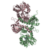
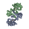
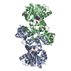
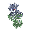
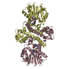
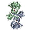
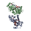
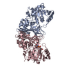
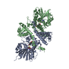
| Title | Crystal Structure Analysis of LaPhzM |
|---|
 Components Components | Methyltransferase |
|---|
 Keywords Keywords | TRANSFERASE / methyltransferase |
|---|
| Function / homology |  Function and homology information Function and homology information
phenazine biosynthetic process / S-adenosylmethionine-dependent methyltransferase activity / O-methyltransferase activity / S-adenosyl-L-methionine binding / Transferases; Transferring one-carbon groups; Methyltransferases / methylation / protein homodimerization activity / identical protein bindingSimilarity search - Function Acetylserotonin O-methyltransferase, dimerisation domain / : / O-methyltransferase domain / O-methyltransferase COMT-type / O-methyltransferase domain / SAM-dependent O-methyltransferase class II-type profile. / Vaccinia Virus protein VP39 / Winged helix-like DNA-binding domain superfamily/Winged helix DNA-binding domain / Arc Repressor Mutant, subunit A / Winged helix DNA-binding domain superfamily ...Acetylserotonin O-methyltransferase, dimerisation domain / : / O-methyltransferase domain / O-methyltransferase COMT-type / O-methyltransferase domain / SAM-dependent O-methyltransferase class II-type profile. / Vaccinia Virus protein VP39 / Winged helix-like DNA-binding domain superfamily/Winged helix DNA-binding domain / Arc Repressor Mutant, subunit A / Winged helix DNA-binding domain superfamily / Winged helix-like DNA-binding domain superfamily / S-adenosyl-L-methionine-dependent methyltransferase superfamily / Rossmann fold / Orthogonal Bundle / 3-Layer(aba) Sandwich / Mainly Alpha / Alpha BetaSimilarity search - Domain/homology |
|---|
| Biological species |  Lysobacter antibioticus (bacteria) Lysobacter antibioticus (bacteria) |
|---|
| Method |  X-RAY DIFFRACTION / SYNCHROTRON / MOLECULAR REPLACEMENT / molecular replacement / Resolution: 1.42 Å X-RAY DIFFRACTION / SYNCHROTRON / MOLECULAR REPLACEMENT / molecular replacement / Resolution: 1.42 Å |
|---|
 Authors Authors | Beltran, D.G. / Schacht, A. / Zhang, L. |
|---|
 Citation Citation | Journal: ACS Chem. Biol. / Year: 2018
Title: Functional and Structural Analysis of Phenazine O-Methyltransferase LaPhzM from Lysobacter antibioticus OH13 and One-Pot Enzymatic Synthesis of the Antibiotic Myxin.
Authors: Jiang, J. / Guiza Beltran, D. / Schacht, A. / Wright, S. / Zhang, L. / Du, L. |
|---|
| History | | Deposition | Jan 15, 2018 | Deposition site: RCSB / Processing site: RCSB |
|---|
| Revision 1.0 | Mar 21, 2018 | Provider: repository / Type: Initial release |
|---|
| Revision 1.1 | May 2, 2018 | Group: Data collection / Database references / Category: citation
Item: _citation.journal_volume / _citation.page_first / _citation.page_last |
|---|
| Revision 2.0 | Oct 4, 2023 | Group: Atomic model / Data collection ...Atomic model / Data collection / Database references / Derived calculations / Other / Refinement description / Structure summary
Category: atom_site / atom_sites ...atom_site / atom_sites / chem_comp / chem_comp_atom / chem_comp_bond / database_2 / entity / pdbx_initial_refinement_model / pdbx_nonpoly_scheme / pdbx_struct_conn_angle / refine / refine_hist / refine_ls_restr / refine_ls_shell / reflns / reflns_shell / struct_conn / struct_site / struct_site_gen
Item: _atom_site.auth_seq_id / _atom_sites.fract_transf_matrix[2][1] ..._atom_site.auth_seq_id / _atom_sites.fract_transf_matrix[2][1] / _atom_sites.fract_transf_matrix[3][2] / _chem_comp.pdbx_synonyms / _database_2.pdbx_DOI / _database_2.pdbx_database_accession / _entity.pdbx_number_of_molecules / _pdbx_struct_conn_angle.ptnr1_auth_asym_id / _pdbx_struct_conn_angle.ptnr1_auth_comp_id / _pdbx_struct_conn_angle.ptnr1_auth_seq_id / _pdbx_struct_conn_angle.ptnr1_label_asym_id / _pdbx_struct_conn_angle.ptnr1_label_comp_id / _pdbx_struct_conn_angle.ptnr1_label_seq_id / _pdbx_struct_conn_angle.ptnr3_auth_asym_id / _pdbx_struct_conn_angle.ptnr3_auth_comp_id / _pdbx_struct_conn_angle.ptnr3_auth_seq_id / _pdbx_struct_conn_angle.ptnr3_label_asym_id / _pdbx_struct_conn_angle.ptnr3_label_comp_id / _pdbx_struct_conn_angle.ptnr3_label_seq_id / _pdbx_struct_conn_angle.value / _refine.B_iso_max / _refine.B_iso_mean / _refine.B_iso_min / _refine.aniso_B[1][2] / _refine.details / _refine.ls_R_factor_R_free / _refine.ls_R_factor_obs / _refine.ls_percent_reflns_obs / _refine.pdbx_starting_model / _refine_hist.cycle_id / _refine_hist.number_atoms_solvent / _refine_hist.number_atoms_total / _refine_hist.pdbx_B_iso_mean_ligand / _refine_hist.pdbx_B_iso_mean_solvent / _refine_hist.pdbx_number_residues_total / _refine_ls_shell.R_factor_R_free_error / _refine_ls_shell.d_res_low / _refine_ls_shell.number_reflns_all / _reflns.number_all / _reflns.pdbx_Rpim_I_all / _reflns.pdbx_Rrim_I_all / _reflns.pdbx_netI_over_av_sigmaI / _reflns.pdbx_number_measured_all / _struct_conn.pdbx_dist_value / _struct_conn.ptnr1_auth_asym_id / _struct_conn.ptnr1_auth_comp_id / _struct_conn.ptnr1_auth_seq_id / _struct_conn.ptnr1_label_asym_id / _struct_conn.ptnr1_label_atom_id / _struct_conn.ptnr1_label_comp_id / _struct_conn.ptnr1_label_seq_id / _struct_conn.ptnr2_auth_asym_id / _struct_conn.ptnr2_auth_comp_id / _struct_conn.ptnr2_auth_seq_id / _struct_conn.ptnr2_label_asym_id / _struct_conn.ptnr2_label_atom_id / _struct_conn.ptnr2_label_comp_id / _struct_conn.ptnr2_label_seq_id |
|---|
|
|---|
 Movie
Movie Controller


 Open data
 Basic information
 Structure visualization
 Downloads & links
 Other downloads
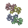

 PDBj
PDBj Assembly
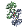


 Type: L-peptide linking / Mass: 384.411 Da / Num. of mol.: 2 / Source method: obtained synthetically / Formula: C14H20N6O5S
 Mass: 46.025 Da / Num. of mol.: 3 / Source method: obtained synthetically / Formula: CH2O2
 Mass: 22.990 Da / Num. of mol.: 3 / Source method: obtained synthetically / Formula: Na
 Mass: 326.383 Da / Num. of mol.: 1 / Source method: obtained synthetically / Formula: C14H30O8 / Comment: precipitant*YM
 Sample preparation
 / Beamline: BL9-2 / Wavelength: 0.97946 Å
 Processing