National Institutes of Health/National Institute of General Medical Sciences (NIH/NIGMS)
GM122510
United States
Citation
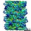
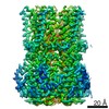
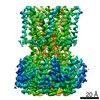
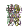
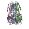
Journal: Elife / Year: 2018 Title: Functional role of the type 1 pilus rod structure in mediating host-pathogen interactions. Authors: Caitlin N Spaulding / Henry Louis Schreiber / Weili Zheng / Karen W Dodson / Jennie E Hazen / Matt S Conover / Fengbin Wang / Pontus Svenmarker / Areli Luna-Rico / Olivera Francetic / Magnus ...Authors: Caitlin N Spaulding / Henry Louis Schreiber / Weili Zheng / Karen W Dodson / Jennie E Hazen / Matt S Conover / Fengbin Wang / Pontus Svenmarker / Areli Luna-Rico / Olivera Francetic / Magnus Andersson / Scott Hultgren / Edward H Egelman / Abstract: Uropathogenic (UPEC), which cause urinary tract infections (UTI), utilize type 1 pili, a chaperone usher pathway (CUP) pilus, to cause UTI and colonize the gut. The pilus rod, comprised of repeating ...Uropathogenic (UPEC), which cause urinary tract infections (UTI), utilize type 1 pili, a chaperone usher pathway (CUP) pilus, to cause UTI and colonize the gut. The pilus rod, comprised of repeating FimA subunits, provides a structural scaffold for displaying the tip adhesin, FimH. We solved the 4.2 Å resolution structure of the type 1 pilus rod using cryo-electron microscopy. Residues forming the interactive surfaces that determine the mechanical properties of the rod were maintained by selection based on a global alignment of sequences. We identified mutations that did not alter pilus production in vitro but reduced the force required to unwind the rod. UPEC expressing these mutant pili were significantly attenuated in bladder infection and intestinal colonization in mice. This study elucidates an unappreciated functional role for the molecular spring-like property of type 1 pilus rods in host-pathogen interactions and carries important implications for other pilus-mediated diseases.
Helical symmetry: (Circular symmetry: 1 / Dyad axis: no / N subunits divisor: 1 / Num. of operations: 10 / Rise per n subunits: 7.7 Å / Rotation per n subunits: 115 °)
Details
THE HELICAL SYMMETRY PARAMETER SHOULD BE APPLIED TO A SINGLE CHAIN OF THE MODEL, INSTEAD OF WHOLE MODEL IN COORDINATES FILE, TO ACHIEVE THE ENTIRE ASSEMBLY.
-
Components
#1: Protein
Type-1fimbrialprotein, Achain / Type-1A pilin
Mass: 15835.243 Da / Num. of mol.: 11 / Source method: isolated from a natural source / Source: (natural) Escherichia coli (E. coli) / Strain: K12 / References: UniProt: P04128
Has protein modification
Y
-
Experimental details
-
Experiment
Experiment
Method: ELECTRON MICROSCOPY
EM experiment
Aggregation state: FILAMENT / 3D reconstruction method: helical reconstruction
-
Sample preparation
Component
Name: Type 1 pilus / Type: COMPLEX / Entity ID: all / Source: NATURAL
Molecular weight
Experimental value: NO
Source (natural)
Organism: Escherichia coli K-12 (bacteria)
Buffer solution
pH: 7.4
Specimen
Embedding applied: NO / Shadowing applied: NO / Staining applied: NO / Vitrification applied: YES
Specimen support
Details: unspecified
Vitrification
Cryogen name: ETHANE / Humidity: 95 %
-
Electron microscopy imaging
Experimental equipment
Model: Titan Krios / Image courtesy: FEI Company
Microscopy
Model: FEI TITAN KRIOS
Electron gun
Electron source: FIELD EMISSION GUN / Accelerating voltage: 300 kV / Illumination mode: FLOOD BEAM
Electron lens
Mode: BRIGHT FIELD
Image recording
Electron dose: 20 e/Å2 / Detector mode: INTEGRATING / Film or detector model: FEI FALCON II (4k x 4k)
In the structure databanks used in Yorodumi, some data are registered as the other names, "COVID-19 virus" and "2019-nCoV". Here are the details of the virus and the list of structure data.
Jan 31, 2019. EMDB accession codes are about to change! (news from PDBe EMDB page)
EMDB accession codes are about to change! (news from PDBe EMDB page)
The allocation of 4 digits for EMDB accession codes will soon come to an end. Whilst these codes will remain in use, new EMDB accession codes will include an additional digit and will expand incrementally as the available range of codes is exhausted. The current 4-digit format prefixed with “EMD-” (i.e. EMD-XXXX) will advance to a 5-digit format (i.e. EMD-XXXXX), and so on. It is currently estimated that the 4-digit codes will be depleted around Spring 2019, at which point the 5-digit format will come into force.
The EM Navigator/Yorodumi systems omit the EMD- prefix.
Related info.:Q: What is EMD? / ID/Accession-code notation in Yorodumi/EM Navigator
Yorodumi is a browser for structure data from EMDB, PDB, SASBDB, etc.
This page is also the successor to EM Navigator detail page, and also detail information page/front-end page for Omokage search.
The word "yorodu" (or yorozu) is an old Japanese word meaning "ten thousand". "mi" (miru) is to see.
Related info.:EMDB / PDB / SASBDB / Comparison of 3 databanks / Yorodumi Search / Aug 31, 2016. New EM Navigator & Yorodumi / Yorodumi Papers / Jmol/JSmol / Function and homology information / Changes in new EM Navigator and Yorodumi
 Movie
Movie Controller


 Open data
 Basic information
 Components
 Keywords
 Function and homology information
 Authors
 United States, 1items
  Citation

 Structure visualization
 Downloads & links
 Other downloads
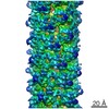

 PDBj
PDBj Assembly
 Sample preparation
 Electron microscopy imaging
 FIELD EMISSION GUN / Accelerating voltage: 300 kV / Illumination mode: FLOOD BEAM
 Processing