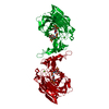
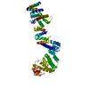
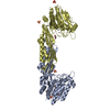
[English] 日本語
Yorodumi
- PDB-6bxb: Crystal structure of an extended b3 integrin P33 -

+
Open data


ID or keywords:

Loading...

-
Basic information

Entry
Database: PDB / ID: 6bxb
TitleCrystal structure of an extended b3 integrin P33
ComponentsChimera protein of Integrin beta-3 and Integrin alpha-L
KeywordsCELL ADHESION / Integrin
Function / homology
Function and homology information


memory T cell extravasation / integrin alphaL-beta2 complex / T cell activation via T cell receptor contact with antigen bound to MHC molecule on antigen presenting cell / ICAM-3 receptor activity / tube development / regulation of serotonin uptake / positive regulation of adenylate cyclase-inhibiting opioid receptor signaling pathway / alpha9-beta1 integrin-ADAM8 complex / regulation of trophoblast cell migration / regulation of postsynaptic neurotransmitter receptor diffusion trapping ...memory T cell extravasation / integrin alphaL-beta2 complex / T cell activation via T cell receptor contact with antigen bound to MHC molecule on antigen presenting cell / ICAM-3 receptor activity / tube development / regulation of serotonin uptake / positive regulation of adenylate cyclase-inhibiting opioid receptor signaling pathway / alpha9-beta1 integrin-ADAM8 complex / regulation of trophoblast cell migration / regulation of postsynaptic neurotransmitter receptor diffusion trapping / alphav-beta3 integrin-vitronectin complex / regulation of extracellular matrix organization / platelet alpha granule membrane / positive regulation of glomerular mesangial cell proliferation / integrin alphav-beta3 complex / negative regulation of lipoprotein metabolic process / alphav-beta3 integrin-PKCalpha complex / fibrinogen binding / maintenance of postsynaptic specialization structure / alphav-beta3 integrin-HMGB1 complex / blood coagulation, fibrin clot formation / negative regulation of lipid transport / vascular endothelial growth factor receptor 2 binding / glycinergic synapse / negative regulation of low-density lipoprotein receptor activity / angiogenesis involved in wound healing / Elastic fibre formation / regulation of release of sequestered calcium ion into cytosol / RUNX3 Regulates Immune Response and Cell Migration / mesodermal cell differentiation / cell-substrate junction assembly / alphav-beta3 integrin-IGF-1-IGF1R complex / platelet-derived growth factor receptor binding / filopodium membrane / extracellular matrix binding / regulation of postsynaptic neurotransmitter receptor internalization / positive regulation of fibroblast migration / positive regulation of vascular endothelial growth factor receptor signaling pathway / apolipoprotein A-I-mediated signaling pathway / regulation of bone resorption / apoptotic cell clearance / positive regulation of cell adhesion mediated by integrin / wound healing, spreading of epidermal cells / heterotypic cell-cell adhesion / integrin complex / Molecules associated with elastic fibres / positive regulation of cell-matrix adhesion / cellular response to insulin-like growth factor stimulus / smooth muscle cell migration / heterophilic cell-cell adhesion via plasma membrane cell adhesion molecules / microvillus membrane / negative chemotaxis / cell adhesion mediated by integrin / leukocyte cell-cell adhesion / Syndecan interactions / p130Cas linkage to MAPK signaling for integrins / activation of protein kinase activity / cellular response to platelet-derived growth factor stimulus / cell-substrate adhesion / protein disulfide isomerase activity / positive regulation of smooth muscle cell migration / positive regulation of osteoblast proliferation / receptor clustering / TGF-beta receptor signaling activates SMADs / PECAM1 interactions / lamellipodium membrane / GRB2:SOS provides linkage to MAPK signaling for Integrins / negative regulation of macrophage derived foam cell differentiation / platelet-derived growth factor receptor signaling pathway / negative regulation of lipid storage / fibronectin binding / ECM proteoglycans / positive regulation of T cell migration / positive regulation of bone resorption / Integrin cell surface interactions / coreceptor activity / specific granule membrane / phagocytosis / negative regulation of endothelial cell apoptotic process / positive regulation of substrate adhesion-dependent cell spreading / cell adhesion molecule binding / positive regulation of endothelial cell proliferation / embryo implantation / positive regulation of endothelial cell migration / Integrin signaling / substrate adhesion-dependent cell spreading / cell-matrix adhesion / response to activity / Signal transduction by L1 / integrin-mediated signaling pathway / Cell surface interactions at the vascular wall / regulation of actin cytoskeleton organization / protein kinase C binding / positive regulation of smooth muscle cell proliferation / Signaling by high-kinase activity BRAF mutants / wound healing / MAP2K and MAPK activation / platelet activation / platelet aggregation / ruffle membrane
Similarity search - Function
Integrin beta, epidermal growth factor-like domain 1 / Integrin beta epidermal growth factor like domain 1 / Integrin beta subunit, cytoplasmic domain / Integrin beta cytoplasmic domain / Integrin_b_cyt / Integrin beta tail domain / Integrin beta subunit, tail / Integrin beta tail domain superfamily / Integrin_B_tail / Integrin beta subunit, VWA domain ...Integrin beta, epidermal growth factor-like domain 1 / Integrin beta epidermal growth factor like domain 1 / Integrin beta subunit, cytoplasmic domain / Integrin beta cytoplasmic domain / Integrin_b_cyt / Integrin beta tail domain / Integrin beta subunit, tail / Integrin beta tail domain superfamily / Integrin_B_tail / Integrin beta subunit, VWA domain / Integrin beta subunit / Integrin beta N-terminal / Integrin beta chain VWA domain / Integrin plexin domain / Integrins beta chain cysteine-rich domain signature. / Integrin beta subunits (N-terminal portion of extracellular region) / Integrin alpha cytoplasmic region / EGF-like domain, extracellular / EGF-like domain / Integrin alpha-2 / Integrin alpha Ig-like domain 1 / : / Integrin alpha Ig-like domain 2 / Integrin alpha chain / Integrin alpha beta-propellor / Integrin alpha chain, C-terminal cytoplasmic region, conserved site / Integrins alpha chain signature. / FG-GAP repeat profile. / Integrin alpha (beta-propellor repeats). / FG-GAP repeat / FG-GAP repeat / Integrin alpha, N-terminal / Integrin domain superfamily / von Willebrand factor type A domain / PSI domain / domain found in Plexins, Semaphorins and Integrins / von Willebrand factor (vWF) type A domain / VWFA domain profile. / von Willebrand factor, type A / von Willebrand factor A-like domain superfamily / EGF-like domain signature 2. / EGF-like domain signature 1.
Similarity search - Domain/homology
Integrin beta-3 / Integrin alpha-L
Similarity search - Component
Biological speciesHomo sapiens (human)
MethodX-RAY DIFFRACTION / SYNCHROTRON / Resolution: 2.39 Å
AuthorsZhou, D. / Zhu, J.
Funding support United States, 1items
OrganizationGrant numberCountry
National Institutes of Health/National Heart, Lung, and Blood Institute (NIH/NHLBI)R01HL131386 United States
CitationJournal: Blood / Year: 2018
Title: Structure of an extended beta3integrin.
Authors: Zhou, D. / Thinn, A.M.M. / Zhao, Y. / Wang, Z. / Zhu, J.
History
DepositionDec 18, 2017Deposition site: RCSB / Processing site: RCSB
Revision 1.0Aug 1, 2018Provider: repository / Type: Initial release
Revision 1.1Sep 12, 2018Group: Data collection / Database references / Category: citation
Item: _citation.journal_volume / _citation.page_first / _citation.page_last
Revision 1.2Dec 4, 2019Group: Author supporting evidence / Category: pdbx_audit_support / Item: _pdbx_audit_support.funding_organization
Revision 1.3Jul 29, 2020Group: Data collection / Derived calculations / Structure summary
Category: chem_comp / entity ...chem_comp / entity / pdbx_chem_comp_identifier / pdbx_entity_nonpoly / pdbx_struct_conn_angle / struct_conn / struct_conn_type / struct_site / struct_site_gen
Item: _chem_comp.name / _chem_comp.type ..._chem_comp.name / _chem_comp.type / _entity.pdbx_description / _pdbx_entity_nonpoly.name / _pdbx_struct_conn_angle.ptnr1_auth_seq_id / _pdbx_struct_conn_angle.ptnr1_symmetry / _pdbx_struct_conn_angle.ptnr3_auth_seq_id / _pdbx_struct_conn_angle.ptnr3_symmetry / _pdbx_struct_conn_angle.value / _struct_conn.conn_type_id / _struct_conn.id / _struct_conn.pdbx_dist_value / _struct_conn.pdbx_leaving_atom_flag / _struct_conn.pdbx_role / _struct_conn.ptnr1_auth_asym_id / _struct_conn.ptnr1_auth_comp_id / _struct_conn.ptnr1_auth_seq_id / _struct_conn.ptnr1_label_asym_id / _struct_conn.ptnr1_label_atom_id / _struct_conn.ptnr1_label_comp_id / _struct_conn.ptnr1_label_seq_id / _struct_conn.ptnr2_auth_asym_id / _struct_conn.ptnr2_auth_comp_id / _struct_conn.ptnr2_auth_seq_id / _struct_conn.ptnr2_label_asym_id / _struct_conn.ptnr2_label_atom_id / _struct_conn.ptnr2_label_comp_id / _struct_conn.ptnr2_symmetry / _struct_conn_type.id
Description: Carbohydrate remediation / Provider: repository / Type: Remediation

-
Structure visualization

Structure viewerMolecule:
MolmilJmol/JSmol

Downloads & links

-
Assembly

Deposited unit
A: Chimera protein of Integrin beta-3 and Integrin alpha-L
B: Chimera protein of Integrin beta-3 and Integrin alpha-L
hetero molecules


Theoretical massNumber of molelcules
Total (without water)105,26011
Polymers103,9052
Non-polymers1,3559
Water3,063170
1
A: Chimera protein of Integrin beta-3 and Integrin alpha-L
hetero molecules


Theoretical massNumber of molelcules
Total (without water)52,6196
Polymers51,9531
Non-polymers6675
Water181
TypeNameSymmetry operationNumber
identity operation1_555x,y,z1
MethodPISA
2
B: Chimera protein of Integrin beta-3 and Integrin alpha-L
hetero molecules


Theoretical massNumber of molelcules
Total (without water)52,6415
Polymers51,9531
Non-polymers6884
Water181
TypeNameSymmetry operationNumber
identity operation1_555x,y,z1
MethodPISA
Unit cell
Length a, b, c (Å)59.560, 79.840, 116.160
Angle α, β, γ (deg.)90.00, 91.42, 90.00
Int Tables number4
Space group name H-MP1211

-
Components

-
Protein / Sugars , 2 types, 7 molecules AB

#1: Protein Chimera protein of Integrin beta-3 and Integrin alpha-L / Platelet membrane glycoprotein IIIa / GPIIIa / CD11 antigen-like family member A / Leukocyte ...Platelet membrane glycoprotein IIIa / GPIIIa / CD11 antigen-like family member A / Leukocyte adhesion glycoprotein LFA-1 alpha chain / LFA-1A / Leukocyte function-associated molecule 1 alpha chain


Mass: 51952.691 Da / Num. of mol.: 2
Source method: isolated from a genetically manipulated source
Source: (gene. exp.) Homo sapiens (human) / Gene: ITGB3, GP3A, ITGAL, CD11A / Production host: Homo sapiens (human) / References: UniProt: P05106, UniProt: P20701
#3: Sugar
ChemComp-NAG / 2-acetamido-2-deoxy-beta-D-glucopyranose


Type: D-saccharide, beta linking / Mass: 221.208 Da / Num. of mol.: 5 / Source method: obtained synthetically / Formula: C8H15NO6
IdentifierTypeProgram
DGlcpNAcbCONDENSED IUPAC CARBOHYDRATE SYMBOLGMML 1.0
N-acetyl-b-D-glucopyranosamineCOMMON NAMEGMML 1.0
b-D-GlcpNAcIUPAC CARBOHYDRATE SYMBOLPDB-CARE 1.0
GlcNAcSNFG CARBOHYDRATE SYMBOLGMML 1.0

-
Non-polymers , 4 types, 174 molecules

#2: Chemical ChemComp-CA / CALCIUM ION


Mass: 40.078 Da / Num. of mol.: 1 / Source method: obtained synthetically / Formula: Ca
#4: Chemical ChemComp-GOL / GLYCEROL / GLYCERIN / PROPANE-1,2,3-TRIOL


Mass: 92.094 Da / Num. of mol.: 2 / Source method: obtained synthetically / Formula: C3H8O3
#5: Chemical ChemComp-MG / MAGNESIUM ION


Mass: 24.305 Da / Num. of mol.: 1 / Source method: obtained synthetically / Formula: Mg
#6: Water ChemComp-HOH / water


Mass: 18.015 Da / Num. of mol.: 170 / Source method: isolated from a natural source / Formula: H2O

-
Experimental details

-
Experiment

ExperimentMethod: X-RAY DIFFRACTION / Number of used crystals: 1

-
Sample preparation

CrystalDensity Matthews: 2.66 Å3/Da / Density % sol: 53.71 %
Crystal growTemperature: 277 K / Method: vapor diffusion, hanging drop / Details: 20% PEG 6000, 0.1 M Tris-HCl (pH 8.5)

-
Data collection

DiffractionMean temperature: 100 K
Diffraction sourceSource: SYNCHROTRON / Site: APS / Beamline: 21-ID-F / Wavelength: 0.97872 Å
DetectorType: MAR CCD 130 mm / Detector: CCD / Date: Jul 13, 2016
RadiationProtocol: SINGLE WAVELENGTH / Monochromatic (M) / Laue (L): M / Scattering type: x-ray
Radiation wavelengthWavelength: 0.97872 Å / Relative weight: 1
ReflectionResolution: 2.388→53.52 Å / Num. obs: 43236 / % possible obs: 99.7 % / Redundancy: 7.6 % / Net I/σ(I): 9.6
Reflection shellResolution: 2.39→2.48 Å

-
Processing

Software
NameVersionClassification
PHENIX1.12_2829refinement
MOSFLMdata reduction
SCALAdata scaling
PHASERphasing
RefinementResolution: 2.39→53.52 Å / SU ML: 0.42 / Cross valid method: FREE R-VALUE / σ(F): 1.34 / Phase error: 32.52
RfactorNum. reflection% reflection
Rfree0.2679 2128 4.93 %
Rwork0.2207 --
obs0.2231 43143 99.4 %
Solvent computationShrinkage radii: 0.9 Å / VDW probe radii: 1.11 Å
Refinement stepCycle: LAST / Resolution: 2.39→53.52 Å
ProteinNucleic acidLigandSolventTotal
Num. atoms7211 0 84 170 7465
Refine LS restraints
Refine-IDTypeDev idealNumber
X-RAY DIFFRACTIONf_bond_d0.0047472
X-RAY DIFFRACTIONf_angle_d0.6510074
X-RAY DIFFRACTIONf_dihedral_angle_d16.6464642
X-RAY DIFFRACTIONf_chiral_restr0.0451116
X-RAY DIFFRACTIONf_plane_restr0.0041318
LS refinement shell
Resolution (Å)Rfactor RfreeNum. reflection RfreeRfactor RworkNum. reflection RworkRefine-ID% reflection obs (%)
2.388-2.44350.36461120.33142652X-RAY DIFFRACTION97
2.4435-2.50470.33691210.32052719X-RAY DIFFRACTION99
2.5047-2.57240.32941380.32492725X-RAY DIFFRACTION99
2.5724-2.64810.35381730.31072669X-RAY DIFFRACTION99
2.6481-2.73350.34761610.31192709X-RAY DIFFRACTION99
2.7335-2.83120.3791230.29912750X-RAY DIFFRACTION99
2.8312-2.94460.32851240.29222737X-RAY DIFFRACTION100
2.9446-3.07860.34191440.26782729X-RAY DIFFRACTION100
3.0786-3.24090.38141460.26172722X-RAY DIFFRACTION100
3.2409-3.44390.33351520.23732745X-RAY DIFFRACTION100
3.4439-3.70970.2681340.22132770X-RAY DIFFRACTION100
3.7097-4.08290.25561540.19992706X-RAY DIFFRACTION100
4.0829-4.67340.2291520.17592776X-RAY DIFFRACTION100
4.6734-5.88690.20581650.18352763X-RAY DIFFRACTION100
5.8869-53.53770.21951290.19382843X-RAY DIFFRACTION100
Refinement TLS params.

Method: refined / Refine-ID: X-RAY DIFFRACTION

IDL112)L122)L132)L222)L232)L332)S11 (Å °)S12 (Å °)S13 (Å °)S21 (Å °)S22 (Å °)S23 (Å °)S31 (Å °)S32 (Å °)S33 (Å °)T112)T122)T132)T222)T232)T332)Origin x (Å)Origin y (Å)Origin z (Å)
10.2254-0.0150.0559-0.0005-0.00470.26680.04680.07590.0602-0.00670.0044-0.0178-0.0879-0.014100.4669-0.05090.01290.344-0.0080.454678.945182.3037135.2793
20.3327-0.22950.13380.54370.03690.1827-0.10310.18160.07490.1827-0.15110.1664-0.22650.1-0.15210.4357-0.0822-0.03450.59960.16840.423166.814555.4217186.8524
Refinement TLS group
IDRefine-IDRefine TLS-IDSelection details
1X-RAY DIFFRACTION1(chain A and resseq 1:463)
2X-RAY DIFFRACTION2(chain B and resseq 1:465)

+
About Yorodumi

-
News

-
Feb 9, 2022. New format data for meta-information of EMDB entries

New format data for meta-information of EMDB entries

  • Version 3 of the EMDB header file is now the official format.
  • The previous official version 1.9 will be removed from the archive.

Related info.:EMDB header

External links:wwPDB to switch to version 3 of the EMDB data model

-
Aug 12, 2020. Covid-19 info

Covid-19 info

URL: https://pdbjlvh1.pdbj.org/emnavi/covid19.php

New page: Covid-19 featured information page in EM Navigator.

Related info.:Covid-19 info / Mar 5, 2020. Novel coronavirus structure data

+
Mar 5, 2020. Novel coronavirus structure data

Novel coronavirus structure data

Related info.:Yorodumi Speices / Aug 12, 2020. Covid-19 info

External links:COVID-19 featured content - PDBj / Molecule of the Month (242):Coronavirus Proteases

+
Jan 31, 2019. EMDB accession codes are about to change! (news from PDBe EMDB page)

EMDB accession codes are about to change! (news from PDBe EMDB page)

  • The allocation of 4 digits for EMDB accession codes will soon come to an end. Whilst these codes will remain in use, new EMDB accession codes will include an additional digit and will expand incrementally as the available range of codes is exhausted. The current 4-digit format prefixed with “EMD-” (i.e. EMD-XXXX) will advance to a 5-digit format (i.e. EMD-XXXXX), and so on. It is currently estimated that the 4-digit codes will be depleted around Spring 2019, at which point the 5-digit format will come into force.
  • The EM Navigator/Yorodumi systems omit the EMD- prefix.

Related info.:Q: What is EMD? / ID/Accession-code notation in Yorodumi/EM Navigator

External links:EMDB Accession Codes are Changing Soon! / Contact to PDBj

+
Jul 12, 2017. Major update of PDB

Major update of PDB

  • wwPDB released updated PDB data conforming to the new PDBx/mmCIF dictionary.
  • This is a major update changing the version number from 4 to 5, and with Remediation, in which all the entries are updated.
  • In this update, many items about electron microscopy experimental information are reorganized (e.g. em_software).
  • Now, EM Navigator and Yorodumi are based on the updated data.

External links:wwPDB Remediation / Enriched Model Files Conforming to OneDep Data Standards Now Available in the PDB FTP Archive

-
Yorodumi

Thousand views of thousand structures

  • Yorodumi is a browser for structure data from EMDB, PDB, SASBDB, etc.
  • This page is also the successor to EM Navigator detail page, and also detail information page/front-end page for Omokage search.
  • The word "yorodu" (or yorozu) is an old Japanese word meaning "ten thousand". "mi" (miru) is to see.

Related info.:EMDB / PDB / SASBDB / Comparison of 3 databanks / Yorodumi Search / Aug 31, 2016. New EM Navigator & Yorodumi / Yorodumi Papers / Jmol/JSmol / Function and homology information / Changes in new EM Navigator and Yorodumi

Read more