 Movie
Movie Controller


+ Open data

- Basic information
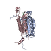
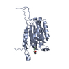
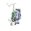
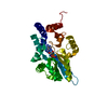
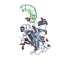
| Entry | Database: PDB / ID: 6bha | ||||||
|---|---|---|---|---|---|---|---|
| Title | Caspase-3 Mutant - T152V | ||||||
 Components Components |
| ||||||
 Keywords Keywords | apoptosis/inhibitor / allosteric regulation / apoptosis / biophysics / caspase / computational biology / X-ray crystallography / fluorescence / molecular dynamics / protein evolution / apoptosis-inhibitor complex | ||||||
| Function / homology |  Function and homology information Function and homology informationcaspase-3 / phospholipase A2 activator activity / Stimulation of the cell death response by PAK-2p34 / anterior neural tube closure / intrinsic apoptotic signaling pathway in response to osmotic stress / leukocyte apoptotic process / glial cell apoptotic process / NADE modulates death signalling / positive regulation of pyroptotic inflammatory response / luteolysis ...caspase-3 / phospholipase A2 activator activity / Stimulation of the cell death response by PAK-2p34 / anterior neural tube closure / intrinsic apoptotic signaling pathway in response to osmotic stress / leukocyte apoptotic process / glial cell apoptotic process / NADE modulates death signalling / positive regulation of pyroptotic inflammatory response / luteolysis / response to cobalt ion / cellular response to staurosporine / cyclin-dependent protein serine/threonine kinase inhibitor activity / death-inducing signaling complex / Apoptotic cleavage of cell adhesion proteins / Caspase activation via Dependence Receptors in the absence of ligand / Apoptosis induced DNA fragmentation / SMAC, XIAP-regulated apoptotic response / Activation of caspases through apoptosome-mediated cleavage / platelet formation / axonal fasciculation / Signaling by Hippo / SMAC (DIABLO) binds to IAPs / SMAC(DIABLO)-mediated dissociation of IAP:caspase complexes / epithelial cell apoptotic process / death receptor binding / fibroblast apoptotic process / regulation of synaptic vesicle cycle / Other interleukin signaling / response to anesthetic / execution phase of apoptosis / negative regulation of cytokine production / neurotrophin TRK receptor signaling pathway / positive regulation of amyloid-beta formation / Apoptotic cleavage of cellular proteins / negative regulation of B cell proliferation / negative regulation of activated T cell proliferation / B cell homeostasis / T cell homeostasis / negative regulation of cell cycle / response to amino acid / response to tumor necrosis factor / pyroptotic inflammatory response / Pyroptosis / keratinocyte differentiation / cell fate commitment / regulation of macroautophagy / Caspase-mediated cleavage of cytoskeletal proteins / response to X-ray / response to insulin-like growth factor stimulus / response to glucose / response to UV / swimming behavior / Degradation of the extracellular matrix / striated muscle cell differentiation / intrinsic apoptotic signaling pathway / response to glucocorticoid / protein catabolic process / erythrocyte differentiation / hippocampus development / response to nicotine / apoptotic signaling pathway / response to hydrogen peroxide / sensory perception of sound / regulation of protein stability / protein maturation / protein processing / response to wounding / enzyme activator activity / neuron differentiation / response to estradiol / peptidase activity / positive regulation of neuron apoptotic process / heart development / neuron apoptotic process / protease binding / response to lipopolysaccharide / aspartic-type endopeptidase activity / response to ethanol / learning or memory / response to hypoxia / postsynaptic density / response to xenobiotic stimulus / cysteine-type endopeptidase activity / neuronal cell body / apoptotic process / DNA damage response / protein-containing complex binding / glutamatergic synapse / proteolysis / nucleoplasm / nucleus / cytosol / cytoplasm Similarity search - Function | ||||||
| Biological species |  Homo sapiens (human) Homo sapiens (human) | ||||||
| Method |  X-RAY DIFFRACTION / SYNCHROTRON / Resolution: 1.603 Å X-RAY DIFFRACTION / SYNCHROTRON / Resolution: 1.603 Å | ||||||
 Authors Authors | Thomas, M.E. / Grinshpon, R. / Swartz, P.D. / Clark, A.C. | ||||||
 Citation Citation | Journal: J. Biol. Chem. / Year: 2018 Title: Modifications to a common phosphorylation network provide individualized control in caspases. Authors: Thomas, M.E. / Grinshpon, R. / Swartz, P. / Clark, A.C. | ||||||
| History |
|
- Structure visualization
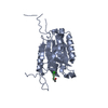
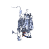
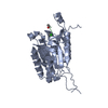
| Structure viewer | Molecule: MolmilJmol/JSmol |
|---|
- Downloads & links
-Download
| PDBx/mmCIF format | 6bha.cif.gz | 71.5 KB | Display | PDBx/mmCIF format |
|---|---|---|---|---|
| PDB format | pdb6bha.ent.gz | 51.9 KB | Display | PDB format |
| PDBx/mmJSON format | 6bha.json.gz | Tree view | PDBx/mmJSON format | |
| Others |  Other downloads Other downloads |
-Validation report
| Arichive directory | https://data.pdbj.org/pub/pdb/validation_reports/bh/6bhaftp://data.pdbj.org/pub/pdb/validation_reports/bh/6bha | HTTPS FTP |
|---|
-Related structure data
| Related structure data |  6bdvC  6bfjC  6bfkC  6bflC  6bfoC  6bg0C 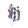 6bg1C  6bg4C  6bgkC  6bgqC  6bgrC  6bgsC  6bh9C C: citing same article ( |
|---|---|
| Similar structure data |

-Links
 PDBj
PDBj


- Assembly
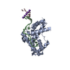
| Deposited unit | 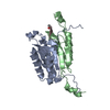
| ||||||||
|---|---|---|---|---|---|---|---|---|---|
| 1 |
| ||||||||
| Unit cell |
|
-Components
| #1: Protein | Mass: 19757.371 Da / Num. of mol.: 1 / Mutation: T152V Source method: isolated from a genetically manipulated source Source: (gene. exp.) Homo sapiens (human) / Gene: CASP3, CPP32 / Production host:  |
|---|---|
| #2: Protein | Mass: 12048.750 Da / Num. of mol.: 1 Source method: isolated from a genetically manipulated source Source: (gene. exp.) Homo sapiens (human) / Gene: CASP3, CPP32 / Production host: |
| #3: Protein/peptide | 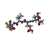  Type: Peptide-like / Class: Inhibitor / Mass: 534.946 Da / Num. of mol.: 1 / Source method: obtained synthetically / Source: (synth.) Homo sapiens (human) / References: Ac-Asp-Glu-Val-Asp-CMK Type: Peptide-like / Class: Inhibitor / Mass: 534.946 Da / Num. of mol.: 1 / Source method: obtained synthetically / Source: (synth.) Homo sapiens (human) / References: Ac-Asp-Glu-Val-Asp-CMK |
| #4: Water | ChemComp-HOH /  Mass: 18.015 Da / Num. of mol.: 203 / Source method: isolated from a natural source / Formula: H2O Mass: 18.015 Da / Num. of mol.: 203 / Source method: isolated from a natural source / Formula: H2O |
| Has protein modification | Y |
-Experimental details
-Experiment
| Experiment | Method: X-RAY DIFFRACTION / Number of used crystals: 1 |
|---|
- Sample preparation
| Crystal | Density Matthews: 2.17 Å3/Da / Density % sol: 43.23 % |
|---|---|
| Crystal grow | Temperature: 291 K / Method: vapor diffusion, hanging drop Details: Crystals were obtained at 18 C by the hanging drop vapor diffusion method using 4 mL drops that contained equal volumes of protein and reservoir solutions over a 0.5 mL solution of 100 mM ...Details: Crystals were obtained at 18 C by the hanging drop vapor diffusion method using 4 mL drops that contained equal volumes of protein and reservoir solutions over a 0.5 mL solution of 100 mM sodium citrate, pH 4.9-5.2, 8-18 % PEG 6000 (w/v), 10 mM DTT, and 3 mM NaN3. Crystals appeared within 3-5 days and were briefly immersed in a cryogenic solution containing 10% MPD (2-methylpentane-2,4-diol) and 90% reservoir solution. |
-Data collection
| Diffraction | Mean temperature: 100 K |
|---|---|
| Diffraction source | Source: SYNCHROTRON / Site: APS  / Beamline: 22-ID / Wavelength: 1 Å / Beamline: 22-ID / Wavelength: 1 Å |
| Detector | Type: MAR scanner 300 mm plate / Detector: IMAGE PLATE / Date: Apr 22, 2016 |
| Radiation | Protocol: SINGLE WAVELENGTH / Monochromatic (M) / Laue (L): M / Scattering type: x-ray |
| Radiation wavelength | Wavelength: 1 Å / Relative weight: 1 |
| Reflection | Resolution: 1.6→50 Å / Num. obs: 36946 / % possible obs: 99.3 % / Redundancy: 7.3 % / Net I/σ(I): 50.4 |
- Processing
| Software |
| |||||||||||||||||||||||||||||||||||||||||||||||||||||||||||||||||||||||||||||||||||||||||||||||||||||||||
|---|---|---|---|---|---|---|---|---|---|---|---|---|---|---|---|---|---|---|---|---|---|---|---|---|---|---|---|---|---|---|---|---|---|---|---|---|---|---|---|---|---|---|---|---|---|---|---|---|---|---|---|---|---|---|---|---|---|---|---|---|---|---|---|---|---|---|---|---|---|---|---|---|---|---|---|---|---|---|---|---|---|---|---|---|---|---|---|---|---|---|---|---|---|---|---|---|---|---|---|---|---|---|---|---|---|---|
| Refinement | Resolution: 1.603→31.794 Å / SU ML: 0.12 / Cross valid method: FREE R-VALUE / σ(F): 1.38 / Phase error: 16.78 Details: Authors indicate that is not possible to define the covalent bond between the cysteine sulfur atom and the carbon atom of the inhibitor in Phenix.
| |||||||||||||||||||||||||||||||||||||||||||||||||||||||||||||||||||||||||||||||||||||||||||||||||||||||||
| Solvent computation | Shrinkage radii: 0.9 Å / VDW probe radii: 1.11 Å | |||||||||||||||||||||||||||||||||||||||||||||||||||||||||||||||||||||||||||||||||||||||||||||||||||||||||
| Refinement step | Cycle: LAST / Resolution: 1.603→31.794 Å
| |||||||||||||||||||||||||||||||||||||||||||||||||||||||||||||||||||||||||||||||||||||||||||||||||||||||||
| Refine LS restraints |
| |||||||||||||||||||||||||||||||||||||||||||||||||||||||||||||||||||||||||||||||||||||||||||||||||||||||||
| LS refinement shell |
|
