 Movie
Movie Controller


+ Open data

- Basic information
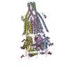
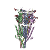
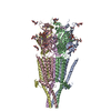
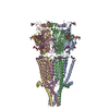
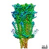
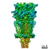
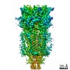
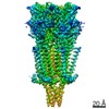
| Entry | Database: PDB / ID: 6be1 | ||||||||||||
|---|---|---|---|---|---|---|---|---|---|---|---|---|---|
| Title | Cryo-EM structure of serotonin receptor | ||||||||||||
 Components Components | 5-hydroxytryptamine receptor 3A | ||||||||||||
 Keywords Keywords | MEMBRANE PROTEIN / Ion Channel | ||||||||||||
| Function / homology |  Function and homology information Function and homology informationNeurotransmitter receptors and postsynaptic signal transmission / serotonin-gated cation-selective signaling pathway / serotonin-activated cation-selective channel complex / serotonin-gated monoatomic cation channel activity / serotonin receptor signaling pathway / serotonin binding / transmembrane transporter complex / excitatory extracellular ligand-gated monoatomic ion channel activity / : / cleavage furrow ...Neurotransmitter receptors and postsynaptic signal transmission / serotonin-gated cation-selective signaling pathway / serotonin-activated cation-selective channel complex / serotonin-gated monoatomic cation channel activity / serotonin receptor signaling pathway / serotonin binding / transmembrane transporter complex / excitatory extracellular ligand-gated monoatomic ion channel activity / : / cleavage furrow / ligand-gated monoatomic ion channel activity involved in regulation of presynaptic membrane potential / transmitter-gated monoatomic ion channel activity involved in regulation of postsynaptic membrane potential / regulation of membrane potential / presynaptic membrane / monoatomic ion transmembrane transport / chemical synaptic transmission / postsynaptic membrane / neuron projection / axon / neuronal cell body / synapse / glutamatergic synapse / identical protein binding / plasma membrane Similarity search - Function | ||||||||||||
| Biological species |  | ||||||||||||
| Method | ELECTRON MICROSCOPY / single particle reconstruction / cryo EM / Resolution: 4.31 Å | ||||||||||||
 Authors Authors | Basak, S. / Chakrapani, S. | ||||||||||||
| Funding support |  United States, 3items United States, 3items
| ||||||||||||
 Citation Citation | Journal: Nat Commun / Year: 2018 Title: Cryo-EM structure of 5-HT receptor in its resting conformation. Authors: Sandip Basak / Yvonne Gicheru / Amrita Samanta / Sudheer Kumar Molugu / Wei Huang / Maria la de Fuente / Taylor Hughes / Derek J Taylor / Marvin T Nieman / Vera Moiseenkova-Bell / Sudha Chakrapani / Abstract: Serotonin receptors (5-HTR) directly regulate gut movement, and drugs that inhibit 5-HTR function are used to control emetic reflexes associated with gastrointestinal pathologies and cancer therapies. ...Serotonin receptors (5-HTR) directly regulate gut movement, and drugs that inhibit 5-HTR function are used to control emetic reflexes associated with gastrointestinal pathologies and cancer therapies. The 5-HTR function involves a finely tuned orchestration of three domain movements that include the ligand-binding domain, the pore domain, and the intracellular domain. Here, we present the structure from the full-length 5-HTR channel in the apo-state determined by single-particle cryo-electron microscopy at a nominal resolution of 4.3 Å. In this conformation, the ligand-binding domain adopts a conformation reminiscent of the unliganded state with the pore domain captured in a closed conformation. In comparison to the 5-HTR crystal structure, the full-length channel in the apo-conformation adopts a more expanded conformation of all the three domains with a characteristic twist that is implicated in gating. | ||||||||||||
| History |
|
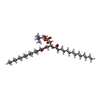
- Structure visualization
| Movie |
Movie viewer |
|---|---|
| Structure viewer | Molecule: MolmilJmol/JSmol |
- Downloads & links
-Download
| PDBx/mmCIF format | 6be1.cif.gz | 551.2 KB | Display | PDBx/mmCIF format |
|---|---|---|---|---|
| PDB format | pdb6be1.ent.gz | 433.4 KB | Display | PDB format |
| PDBx/mmJSON format | 6be1.json.gz | Tree view | PDBx/mmJSON format | |
| Others |  Other downloads Other downloads |
-Validation report
| Arichive directory | https://data.pdbj.org/pub/pdb/validation_reports/be/6be1ftp://data.pdbj.org/pub/pdb/validation_reports/be/6be1 | HTTPS FTP |
|---|
-Related structure data
| Related structure data |  7088MC M: map data used to model this data C: citing same article ( |
|---|---|
| Similar structure data |

-Links
 PDBj
PDBj


- Assembly
| Deposited unit | 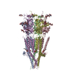
|
|---|---|
| 1 |
|
-Components
-Protein , 1 types, 5 molecules ABCDE
| #1: Protein | Mass: 52739.852 Da / Num. of mol.: 5 Source method: isolated from a genetically manipulated source Source: (gene. exp.)   Spodoptera frugiperda (fall armyworm) / Strain (production host): sf9 / References: UniProt: E9QLC0, UniProt: P23979*PLUS Spodoptera frugiperda (fall armyworm) / Strain (production host): sf9 / References: UniProt: E9QLC0, UniProt: P23979*PLUS |
|---|
-Sugars , 6 types, 18 molecules 

| #2: Polysaccharide | Source method: isolated from a genetically manipulated source #3: Polysaccharide | 2-acetamido-2-deoxy-beta-D-glucopyranose-(1-4)-2-acetamido-2-deoxy-beta-D-glucopyranose Source method: isolated from a genetically manipulated source #4: Polysaccharide | beta-D-mannopyranose-(1-4)-2-acetamido-2-deoxy-beta-D-glucopyranose-(1-3)-2-acetamido-2-deoxy-beta- ...beta-D-mannopyranose-(1-4)-2-acetamido-2-deoxy-beta-D-glucopyranose-(1-3)-2-acetamido-2-deoxy-beta-D-glucopyranose | Source method: isolated from a genetically manipulated source #5: Polysaccharide | beta-D-mannopyranose-(1-3)-2-acetamido-2-deoxy-beta-D-glucopyranose-(1-4)-2-acetamido-2-deoxy-beta- ...beta-D-mannopyranose-(1-3)-2-acetamido-2-deoxy-beta-D-glucopyranose-(1-4)-2-acetamido-2-deoxy-beta-D-glucopyranose | Source method: isolated from a genetically manipulated source #6: Sugar | ChemComp-NAG /  Type: D-saccharide, beta linking / Mass: 221.208 Da / Num. of mol.: 5 Type: D-saccharide, beta linking / Mass: 221.208 Da / Num. of mol.: 5Source method: isolated from a genetically manipulated source Formula: C8H15NO6 #10: Sugar | ChemComp-BMA / |  Type: D-saccharide, beta linking / Mass: 180.156 Da / Num. of mol.: 1 Type: D-saccharide, beta linking / Mass: 180.156 Da / Num. of mol.: 1Source method: isolated from a genetically manipulated source Formula: C6H12O6 |
|---|
-Non-polymers , 4 types, 18 molecules 


| #7: Chemical | ChemComp-NA /  Mass: 22.990 Da / Num. of mol.: 1 / Source method: obtained synthetically / Formula: Na Mass: 22.990 Da / Num. of mol.: 1 / Source method: obtained synthetically / Formula: Na | ||||
|---|---|---|---|---|---|
| #8: Chemical | ChemComp-PX4 /  Mass: 678.940 Da / Num. of mol.: 5 / Source method: obtained synthetically / Formula: C36H73NO8P / Comment: DMPC, phospholipid*YM Mass: 678.940 Da / Num. of mol.: 5 / Source method: obtained synthetically / Formula: C36H73NO8P / Comment: DMPC, phospholipid*YM#9: Chemical | ChemComp-CL / |  Mass: 35.453 Da / Num. of mol.: 1 / Source method: obtained synthetically / Formula: Cl Mass: 35.453 Da / Num. of mol.: 1 / Source method: obtained synthetically / Formula: Cl#11: Water | ChemComp-HOH / | Mass: 18.015 Da / Num. of mol.: 11 / Source method: isolated from a natural source / Formula: H2O |
-Details
| Has protein modification | Y |
|---|
-Experimental details
-Experiment
| Experiment | Method: ELECTRON MICROSCOPY |
|---|---|
| EM experiment | Aggregation state: PARTICLE / 3D reconstruction method: single particle reconstruction |
- Sample preparation
| Component | Name: Serotonin receptor / Type: ORGANELLE OR CELLULAR COMPONENT / Entity ID: #1 / Source: RECOMBINANT |
|---|---|
| Molecular weight | Value: 270 kDa/nm / Experimental value: NO |
| Source (natural) | Organism: |
| Source (recombinant) | Organism: Spodoptera frugiperda (fall armyworm) / Strain: sf9 |
| Buffer solution | pH: 8 |
| Specimen | Conc.: 2.5 mg/ml / Embedding applied: NO / Shadowing applied: NO / Staining applied: NO / Vitrification applied: YES |
| Specimen support | Grid material: COPPER / Grid mesh size: 300 divisions/in. / Grid type: Quantifoil R1.2/1.3 |
| Vitrification | Instrument: FEI VITROBOT MARK I / Cryogen name: ETHANE / Humidity: 100 % / Chamber temperature: 277 K / Details: blot for 2.5 sec. |
- Electron microscopy imaging
| Experimental equipment |  Model: Titan Krios / Image courtesy: FEI Company |
|---|---|
| Microscopy | Model: FEI TITAN KRIOS |
| Electron gun | Electron source:  FIELD EMISSION GUN / Accelerating voltage: 300 kV / Illumination mode: FLOOD BEAM FIELD EMISSION GUN / Accelerating voltage: 300 kV / Illumination mode: FLOOD BEAM |
| Electron lens | Mode: BRIGHT FIELD / Nominal magnification: 130000 X / Cs: 2.7 mm / C2 aperture diameter: 70 µm |
| Specimen holder | Cryogen: NITROGEN |
| Image recording | Electron dose: 40 e/Å2 / Detector mode: COUNTING / Film or detector model: GATAN K2 SUMMIT (4k x 4k) / Num. of real images: 3550 |
| Image scans | Movie frames/image: 40 / Used frames/image: 2-39 |
- Processing
| Software | Name: PHENIX / Version: 1.12_2829: / Classification: refinement | ||||||||||||||||||||||||||||||||||||
|---|---|---|---|---|---|---|---|---|---|---|---|---|---|---|---|---|---|---|---|---|---|---|---|---|---|---|---|---|---|---|---|---|---|---|---|---|---|
| EM software |
| ||||||||||||||||||||||||||||||||||||
| CTF correction | Type: PHASE FLIPPING AND AMPLITUDE CORRECTION | ||||||||||||||||||||||||||||||||||||
| Particle selection | Num. of particles selected: 327329 | ||||||||||||||||||||||||||||||||||||
| Symmetry | Point symmetry: C5 (5 fold cyclic) | ||||||||||||||||||||||||||||||||||||
| 3D reconstruction | Resolution: 4.31 Å / Resolution method: FSC 0.143 CUT-OFF / Num. of particles: 108727 / Num. of class averages: 2 / Symmetry type: 3D CRYSTAL | ||||||||||||||||||||||||||||||||||||
| Atomic model building | B value: 238 / Protocol: OTHER / Space: REAL | ||||||||||||||||||||||||||||||||||||
| Refine LS restraints |
|
