- PDB-6b9q: Single particle cryo-EM structure determination of the LuIII caps... -
+
Open data
ID or keywords:
Loading...
-
Basic information
Entry
Database: PDB / ID: 6b9q
Title
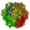
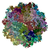
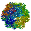
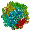
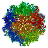
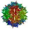
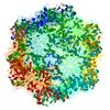
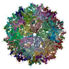
Single particle cryo-EM structure determination of the LuIII capsid protein
Components
Capsid protein VP2
Keywords
VIRUS LIKE PARTICLE / Parvoviridae / VP2 capsid protein
Function / homology
Function and homology information
symbiont entry into host cell via permeabilization of host membrane / microtubule-dependent intracellular transport of viral material towards nucleus / T=1 icosahedral viral capsid / viral penetration into host nucleus / host cell / clathrin-dependent endocytosis of virus by host cell / virion attachment to host cell / host cell nucleus / structural molecule activity / metal ion binding Similarity search - Function
National Institutes of Health/National Institute of General Medical Sciences (NIH/NIGMS)
GM082946-07S1, 229
United States
National Institutes of Health/National Institute of General Medical Sciences (NIH/NIGMS)
GM082946
United States
National Institutes of Health/National Institute Of Allergy and Infectious Diseases (NIH/NIAID)
5T32AI007110-34
United States
National Institutes of Health/National Cancer Institute (NIH/NCI)
CA029303
United States
National Institutes of Health/National Institute of General Medical Sciences (NIH/NIGMS)
U24GM116792
United States
Citation
Journal: Viruses / Year: 2017 Title: Atomic Resolution Structure of the Oncolytic Parvovirus LuIII by Electron Microscopy and 3D Image Reconstruction. Authors: Nikéa Pittman / Adam Misseldine / Lorena Geilen / Sujata Halder / J Kennon Smith / Justin Kurian / Paul Chipman / Mandy Janssen / Robert Mckenna / Timothy S Baker / Anthony D'Abramo / Susan ...Authors: Nikéa Pittman / Adam Misseldine / Lorena Geilen / Sujata Halder / J Kennon Smith / Justin Kurian / Paul Chipman / Mandy Janssen / Robert Mckenna / Timothy S Baker / Anthony D'Abramo / Susan Cotmore / Peter Tattersall / Mavis Agbandje-McKenna / Abstract: LuIII, a protoparvovirus pathogenic to rodents, replicates in human mitotic cells, making it applicable for use to kill cancer cells. This virus group includes H-1 parvovirus (H-1PV) and minute virus ...LuIII, a protoparvovirus pathogenic to rodents, replicates in human mitotic cells, making it applicable for use to kill cancer cells. This virus group includes H-1 parvovirus (H-1PV) and minute virus of mice (MVM). However, LuIII displays enhanced oncolysis compared to H-1PV and MVM, a phenotype mapped to the major capsid viral protein 2 (VP2). This suggests that within LuIII VP2 are determinants for improved tumor lysis. To investigate this, the structure of the LuIII virus-like-particle was determined using single particle cryo-electron microscopy and image reconstruction to 3.17 Å resolution, and compared to the H-1PV and MVM structures. The LuIII VP2 structure, ordered from residue 37 to 587 (C-terminal), had the conserved VP topology and capsid morphology previously reported for other protoparvoviruses. This includes a core β-barrel and α-helix A, a depression at the icosahedral 2-fold and surrounding the 5-fold axes, and a single protrusion at the 3-fold axes. Comparative analysis identified surface loop differences among LuIII, H-1PV, and MVM at or close to the capsid 2- and 5-fold symmetry axes, and the shoulder of the 3-fold protrusions. The 2-fold differences cluster near the previously identified MVM sialic acid receptor binding pocket, and revealed potential determinants of protoparvovirus tumor tropism.
History
Deposition
Oct 11, 2017
Deposition site: RCSB / Processing site: RCSB
Revision 1.0
Nov 15, 2017
Provider: repository / Type: Initial release
Revision 1.0
Nov 15, 2017
Data content type: EM metadata / Data content type: EM metadata / Provider: repository / Type: Initial release
Revision 1.0
Nov 15, 2017
Data content type: Image / Data content type: Image / Provider: repository / Type: Initial release
Revision 1.0
Nov 15, 2017
Data content type: Primary map / Data content type: Primary map / Provider: repository / Type: Initial release
Revision 1.0
Nov 15, 2017
Data content type: Image / Data content type: Image / Provider: repository / Type: Initial release
Revision 1.0
Nov 15, 2017
Data content type: Primary map / Data content type: Primary map / Provider: repository / Type: Initial release
Revision 1.0
Nov 15, 2017
Data content type: Image / Data content type: Image / Provider: repository / Type: Initial release
Revision 1.0
Nov 15, 2017
Data content type: Primary map / Data content type: Primary map / Provider: repository / Type: Initial release
Revision 1.0
Nov 15, 2017
Data content type: Image / Data content type: Image / Provider: repository / Type: Initial release
Revision 1.0
Nov 15, 2017
Data content type: Primary map / Data content type: Primary map / Provider: repository / Type: Initial release
Data content type: EM metadata / Data content type: EM metadata / EM metadata / Group: Data processing / Experimental summary / Data content type: EM metadata / EM metadata / Category: em_admin / em_software / Data content type: EM metadata / EM metadata / Item: _em_admin.last_update / _em_software.name
#200 - Aug 2016 Quasisymmetry in Icosahedral Viruses similarity (1)
-
Assembly
Deposited unit
A: Capsid protein VP2 B: Capsid protein VP2 C: Capsid protein VP2 D: Capsid protein VP2 E: Capsid protein VP2 F: Capsid protein VP2 G: Capsid protein VP2 H: Capsid protein VP2 I: Capsid protein VP2 J: Capsid protein VP2 K: Capsid protein VP2 L: Capsid protein VP2 M: Capsid protein VP2 N: Capsid protein VP2 O: Capsid protein VP2 P: Capsid protein VP2 Q: Capsid protein VP2 R: Capsid protein VP2 S: Capsid protein VP2 T: Capsid protein VP2 U: Capsid protein VP2 V: Capsid protein VP2 W: Capsid protein VP2 X: Capsid protein VP2 Y: Capsid protein VP2 Z: Capsid protein VP2 1: Capsid protein VP2 2: Capsid protein VP2 3: Capsid protein VP2 4: Capsid protein VP2 5: Capsid protein VP2 6: Capsid protein VP2 a: Capsid protein VP2 b: Capsid protein VP2 c: Capsid protein VP2 d: Capsid protein VP2 e: Capsid protein VP2 f: Capsid protein VP2 g: Capsid protein VP2 h: Capsid protein VP2 i: Capsid protein VP2 j: Capsid protein VP2 k: Capsid protein VP2 l: Capsid protein VP2 m: Capsid protein VP2 n: Capsid protein VP2 o: Capsid protein VP2 p: Capsid protein VP2 q: Capsid protein VP2 r: Capsid protein VP2 s: Capsid protein VP2 t: Capsid protein VP2 u: Capsid protein VP2 v: Capsid protein VP2 w: Capsid protein VP2 x: Capsid protein VP2 y: Capsid protein VP2 z: Capsid protein VP2 7: Capsid protein VP2 8: Capsid protein VP2
Conc.: 0.75 mg/ml / Embedding applied: NO / Shadowing applied: NO / Staining applied: NO / Vitrification applied: YES / Details: This sample was monodisperse.
In the structure databanks used in Yorodumi, some data are registered as the other names, "COVID-19 virus" and "2019-nCoV". Here are the details of the virus and the list of structure data.
Jan 31, 2019. EMDB accession codes are about to change! (news from PDBe EMDB page)
EMDB accession codes are about to change! (news from PDBe EMDB page)
The allocation of 4 digits for EMDB accession codes will soon come to an end. Whilst these codes will remain in use, new EMDB accession codes will include an additional digit and will expand incrementally as the available range of codes is exhausted. The current 4-digit format prefixed with “EMD-” (i.e. EMD-XXXX) will advance to a 5-digit format (i.e. EMD-XXXXX), and so on. It is currently estimated that the 4-digit codes will be depleted around Spring 2019, at which point the 5-digit format will come into force.
The EM Navigator/Yorodumi systems omit the EMD- prefix.
Related info.:Q: What is EMD? / ID/Accession-code notation in Yorodumi/EM Navigator
Yorodumi is a browser for structure data from EMDB, PDB, SASBDB, etc.
This page is also the successor to EM Navigator detail page, and also detail information page/front-end page for Omokage search.
The word "yorodu" (or yorozu) is an old Japanese word meaning "ten thousand". "mi" (miru) is to see.
Related info.:EMDB / PDB / SASBDB / Comparison of 3 databanks / Yorodumi Search / Aug 31, 2016. New EM Navigator & Yorodumi / Yorodumi Papers / Jmol/JSmol / Function and homology information / Changes in new EM Navigator and Yorodumi
 Movie
Movie Controller

 Yorodumi
 Open data
 Basic information
 Components
 Keywords
 Function and homology information
 Parvovirus LuIII
 Authors
 United States, 5items
  Citation
 Structure visualization
 Downloads & links
 Other downloads


 PDBj
PDBj


 Assembly

 Spodoptera frugiperda (fall armyworm) / References: UniProt: P36310
 Sample preparation
 Electron microscopy imaging
 FIELD EMISSION GUN / Accelerating voltage: 300 kV / Illumination mode: FLOOD BEAM
 Processing