 Movie
Movie Controller

[English] 日本語
 Yorodumi
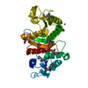
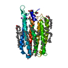
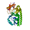
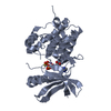
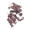
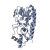
- PDB-6b8v: Crystal structure of adenylyl-sulfate kinase from Cryptococcus ne... -
+ Open data

- Basic information
| Entry | Database: PDB / ID: 6b8v | ||||||
|---|---|---|---|---|---|---|---|
| Title | Crystal structure of adenylyl-sulfate kinase from Cryptococcus neoformans | ||||||
 Components Components | Adenylylsulfate kinase | ||||||
 Keywords Keywords | TRANSFERASE / Seattle Structural Genomics Center for Infectious Disease / SSGCID / Adenylyl-sulfate kinase / hydrogen sulfide biosynthesis | ||||||
| Function / homology |  Function and homology information Function and homology informationadenylyl-sulfate kinase / adenylylsulfate kinase activity / sulfate assimilation / hydrogen sulfide biosynthetic process / L-cysteine biosynthetic process / ATP binding Similarity search - Function | ||||||
| Biological species |  Cryptococcus neoformans (Cryptococcus neoformans serotype A) Cryptococcus neoformans (Cryptococcus neoformans serotype A) | ||||||
| Method |  X-RAY DIFFRACTION / SYNCHROTRON / MOLECULAR REPLACEMENT / Resolution: 2.55 Å X-RAY DIFFRACTION / SYNCHROTRON / MOLECULAR REPLACEMENT / Resolution: 2.55 Å | ||||||
 Authors Authors | Seattle Structural Genomics Center for Infectious Disease (SSGCID) | ||||||
 Citation Citation | Journal: To be Published Title: Crystal structure of adenylyl-sulfate kinase from Cryptococcus neoformans Authors: Delker, S.L. / Abendroth, J. / Lorimer, D. / Edwards, T.E. | ||||||
| History |
|
- Structure visualization
| Structure viewer | Molecule: MolmilJmol/JSmol |
|---|
- Downloads & links
-Download
| PDBx/mmCIF format | 6b8v.cif.gz | 207.7 KB | Display | PDBx/mmCIF format |
|---|---|---|---|---|
| PDB format | pdb6b8v.ent.gz | 165.7 KB | Display | PDB format |
| PDBx/mmJSON format | 6b8v.json.gz | Tree view | PDBx/mmJSON format | |
| Others |  Other downloads Other downloads |
-Validation report
| Arichive directory | https://data.pdbj.org/pub/pdb/validation_reports/b8/6b8vftp://data.pdbj.org/pub/pdb/validation_reports/b8/6b8v | HTTPS FTP |
|---|
-Related structure data
| Related structure data |  2peyS S: Starting model for refinement |
|---|---|
| Similar structure data |

-Links
 PDBj
PDBj
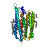
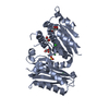
- Assembly
| Deposited unit | 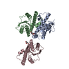
| ||||||||
|---|---|---|---|---|---|---|---|---|---|
| 1 | 
| ||||||||
| 2 | 
| ||||||||
| Unit cell |
| ||||||||
| Components on special symmetry positions |
|
-Components
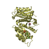
| #1: Protein | Mass: 23297.422 Da / Num. of mol.: 3 / Fragment: CrneC.00830.a.B1 Source method: isolated from a genetically manipulated source Source: (gene. exp.) Cryptococcus neoformans (Cryptococcus neoformans serotype A)Gene: C362_03154 / Variant: grubii serotype A / Plasmid: CrneC.00830.a.B1 / Production host:  #2: Chemical |   Mass: 92.094 Da / Num. of mol.: 2 / Source method: obtained synthetically / Formula: C3H8O3 Mass: 92.094 Da / Num. of mol.: 2 / Source method: obtained synthetically / Formula: C3H8O3#3: Water | ChemComp-HOH / |  Mass: 18.015 Da / Num. of mol.: 139 / Source method: isolated from a natural source / Formula: H2O Mass: 18.015 Da / Num. of mol.: 139 / Source method: isolated from a natural source / Formula: H2O |
|---|
-Experimental details
-Experiment
| Experiment | Method: X-RAY DIFFRACTION / Number of used crystals: 1 |
|---|
- Sample preparation
| Crystal | Density Matthews: 2.76 Å3/Da / Density % sol: 55.38 % |
|---|---|
| Crystal grow | Temperature: 290 K / Method: vapor diffusion, sitting drop / pH: 8.5 Details: 18.6 mg/mL PS38292 against Morpheus A11: 10% w/v PEG4000, 20% v/v glycerol, 0.03 M magnesium chloride, 0.03 M calcium chloride, 0.1 M bicine/Trizma base, pH 8.5, cryoprotection: direct, ...Details: 18.6 mg/mL PS38292 against Morpheus A11: 10% w/v PEG4000, 20% v/v glycerol, 0.03 M magnesium chloride, 0.03 M calcium chloride, 0.1 M bicine/Trizma base, pH 8.5, cryoprotection: direct, CrneC.00830.a.B1, tray 293001, puck ltv3-1 |
-Data collection
| Diffraction | Mean temperature: 100 K | ||||||||||||||||||||||||||||||||||||||||||||||||||||||||||||||||||||||||||||||||||||||||||||||||||||||||||||||||||||||||||||||||||||||||||||||||||||||||||||||||||||||||
|---|---|---|---|---|---|---|---|---|---|---|---|---|---|---|---|---|---|---|---|---|---|---|---|---|---|---|---|---|---|---|---|---|---|---|---|---|---|---|---|---|---|---|---|---|---|---|---|---|---|---|---|---|---|---|---|---|---|---|---|---|---|---|---|---|---|---|---|---|---|---|---|---|---|---|---|---|---|---|---|---|---|---|---|---|---|---|---|---|---|---|---|---|---|---|---|---|---|---|---|---|---|---|---|---|---|---|---|---|---|---|---|---|---|---|---|---|---|---|---|---|---|---|---|---|---|---|---|---|---|---|---|---|---|---|---|---|---|---|---|---|---|---|---|---|---|---|---|---|---|---|---|---|---|---|---|---|---|---|---|---|---|---|---|---|---|---|---|---|---|
| Diffraction source | Source: SYNCHROTRON / Site: APS  / Beamline: 21-ID-F / Wavelength: 0.97872 Å / Beamline: 21-ID-F / Wavelength: 0.97872 Å | ||||||||||||||||||||||||||||||||||||||||||||||||||||||||||||||||||||||||||||||||||||||||||||||||||||||||||||||||||||||||||||||||||||||||||||||||||||||||||||||||||||||||
| Detector | Type: MARMOSAIC 225 mm CCD / Detector: CCD / Date: Aug 4, 2017 / Details: Beryllium Lenses | ||||||||||||||||||||||||||||||||||||||||||||||||||||||||||||||||||||||||||||||||||||||||||||||||||||||||||||||||||||||||||||||||||||||||||||||||||||||||||||||||||||||||
| Radiation | Monochromator: diamond(111) / Protocol: SINGLE WAVELENGTH / Monochromatic (M) / Laue (L): M / Scattering type: x-ray | ||||||||||||||||||||||||||||||||||||||||||||||||||||||||||||||||||||||||||||||||||||||||||||||||||||||||||||||||||||||||||||||||||||||||||||||||||||||||||||||||||||||||
| Radiation wavelength | Wavelength: 0.97872 Å / Relative weight: 1 | ||||||||||||||||||||||||||||||||||||||||||||||||||||||||||||||||||||||||||||||||||||||||||||||||||||||||||||||||||||||||||||||||||||||||||||||||||||||||||||||||||||||||
| Reflection | Resolution: 2.55→43.84 Å / Num. obs: 25257 / % possible obs: 99.8 % / Observed criterion σ(I): -3 / Redundancy: 8.79 % / Biso Wilson estimate: 41.28 Å2 / CC1/2: 0.999 / Rmerge(I) obs: 0.094 / Rrim(I) all: 0.099 / Χ2: 1.05 / Net I/σ(I): 14.76 / Num. measured all: 222005 / Scaling rejects: 10 | ||||||||||||||||||||||||||||||||||||||||||||||||||||||||||||||||||||||||||||||||||||||||||||||||||||||||||||||||||||||||||||||||||||||||||||||||||||||||||||||||||||||||
| Reflection shell | Diffraction-ID: 1
|
- Processing
| Software |
| ||||||||||||||||||||||||||||||||||||||||||||||||||||||||||||||||||||||||||||||||||||||||||||||||||
|---|---|---|---|---|---|---|---|---|---|---|---|---|---|---|---|---|---|---|---|---|---|---|---|---|---|---|---|---|---|---|---|---|---|---|---|---|---|---|---|---|---|---|---|---|---|---|---|---|---|---|---|---|---|---|---|---|---|---|---|---|---|---|---|---|---|---|---|---|---|---|---|---|---|---|---|---|---|---|---|---|---|---|---|---|---|---|---|---|---|---|---|---|---|---|---|---|---|---|---|
| Refinement | Method to determine structure: MOLECULAR REPLACEMENT Starting model: PDB entry 2PEY Resolution: 2.55→43.84 Å / SU ML: 0.3 / Cross valid method: FREE R-VALUE / σ(F): 1.35 / Phase error: 30.47 / Stereochemistry target values: ML
| ||||||||||||||||||||||||||||||||||||||||||||||||||||||||||||||||||||||||||||||||||||||||||||||||||
| Solvent computation | Shrinkage radii: 0.9 Å / VDW probe radii: 1.11 Å / Solvent model: FLAT BULK SOLVENT MODEL | ||||||||||||||||||||||||||||||||||||||||||||||||||||||||||||||||||||||||||||||||||||||||||||||||||
| Refinement step | Cycle: LAST / Resolution: 2.55→43.84 Å
| ||||||||||||||||||||||||||||||||||||||||||||||||||||||||||||||||||||||||||||||||||||||||||||||||||
| Refine LS restraints |
| ||||||||||||||||||||||||||||||||||||||||||||||||||||||||||||||||||||||||||||||||||||||||||||||||||
| LS refinement shell |
| ||||||||||||||||||||||||||||||||||||||||||||||||||||||||||||||||||||||||||||||||||||||||||||||||||
| Refinement TLS params. | Method: refined / Origin x: -0.7226 Å / Origin y: 18.8312 Å / Origin z: -25.3298 Å
| ||||||||||||||||||||||||||||||||||||||||||||||||||||||||||||||||||||||||||||||||||||||||||||||||||
| Refinement TLS group | Selection details: all |
