 Movie
Movie Controller


+ Open data

- Basic information
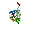
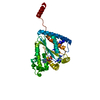
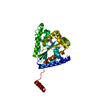
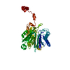
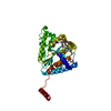
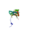
| Entry | Database: PDB / ID: 6b18 | ||||||
|---|---|---|---|---|---|---|---|
| Title | Crystal structure of PPK3 Class III in complex with inhibitor | ||||||
 Components Components | PPK3 Class III | ||||||
 Keywords Keywords | transferase/transferase inhibitor / PPK2 / inhibitor / transferase / transferase-transferase inhibitor complex | ||||||
| Function / homology |  Function and homology information Function and homology informationTransferases; Transferring phosphorus-containing groups; Phosphotransferases with a phosphate group as acceptor / polyphosphate kinase activity / polyphosphate metabolic process Similarity search - Function | ||||||
| Biological species |  Cytophaga hutchinsonii (bacteria) Cytophaga hutchinsonii (bacteria) | ||||||
| Method |  X-RAY DIFFRACTION / SYNCHROTRON / Resolution: 2.3 Å X-RAY DIFFRACTION / SYNCHROTRON / Resolution: 2.3 Å | ||||||
 Authors Authors | Nocek, B. / Ruszkowski, M. / Berlicki, L. / Joachimiak, A. / Yakunin, A. | ||||||
 Citation Citation | Journal: Acs Catalysis / Year: 2018 Title: Structural Insights into Substrate Selectivity and Activity of Bacterial Polyphosphate Kinases Authors: Nocek, B. / Khusnutdinova, A.N. / Ruszkowski, M. / Flick, R. / Burda, M. / Batyrova, K. / Brown, G. / Mucha, A. / Joachimiak, A. / Berlicki, L. / Yakunin, A.F. | ||||||
| History |
|
- Structure visualization
| Structure viewer | Molecule: MolmilJmol/JSmol |
|---|
- Downloads & links
-Download
| PDBx/mmCIF format | 6b18.cif.gz | 141.2 KB | Display | PDBx/mmCIF format |
|---|---|---|---|---|
| PDB format | pdb6b18.ent.gz | 109.8 KB | Display | PDB format |
| PDBx/mmJSON format | 6b18.json.gz | Tree view | PDBx/mmJSON format | |
| Others |  Other downloads Other downloads |
-Validation report
| Arichive directory | https://data.pdbj.org/pub/pdb/validation_reports/b1/6b18ftp://data.pdbj.org/pub/pdb/validation_reports/b1/6b18 | HTTPS FTP |
|---|
-Related structure data
| Related structure data |  6an9C  6angC 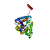 6anhC 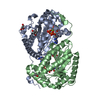 6aqeC  6aqnC  6au0C C: citing same article ( |
|---|---|
| Similar structure data |


-Links
 PDBj
PDBj- Assembly
| Deposited unit | 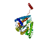
| ||||||||||||
|---|---|---|---|---|---|---|---|---|---|---|---|---|---|
| 1 |
| ||||||||||||
| Unit cell |
| ||||||||||||
| Components on special symmetry positions |
|
-Components
| #1: Protein | Mass: 35577.523 Da / Num. of mol.: 1 Source method: isolated from a genetically manipulated source Source: (gene. exp.) Cytophaga hutchinsonii (bacteria) / Strain: ATCC 33406 / NCIMB 9469Production host: References: UniProt: Q11YW6 |
|---|---|
| #2: Chemical | ChemComp-C8A / {[(  Mass: 357.235 Da / Num. of mol.: 1 / Source method: obtained synthetically / Formula: C14H17NO6P2 / Feature type: SUBJECT OF INVESTIGATION Mass: 357.235 Da / Num. of mol.: 1 / Source method: obtained synthetically / Formula: C14H17NO6P2 / Feature type: SUBJECT OF INVESTIGATION |
| #3: Chemical | ChemComp-PO4 /   Mass: 94.971 Da / Num. of mol.: 1 / Source method: obtained synthetically / Formula: PO4 / Feature type: SUBJECT OF INVESTIGATION Mass: 94.971 Da / Num. of mol.: 1 / Source method: obtained synthetically / Formula: PO4 / Feature type: SUBJECT OF INVESTIGATION |
| #4: Chemical | ChemComp-GOL /   Mass: 92.094 Da / Num. of mol.: 1 / Source method: obtained synthetically / Formula: C3H8O3 / Feature type: SUBJECT OF INVESTIGATION Mass: 92.094 Da / Num. of mol.: 1 / Source method: obtained synthetically / Formula: C3H8O3 / Feature type: SUBJECT OF INVESTIGATION |
| #5: Water | ChemComp-HOH /  Mass: 18.015 Da / Num. of mol.: 174 / Source method: isolated from a natural source / Formula: H2O Mass: 18.015 Da / Num. of mol.: 174 / Source method: isolated from a natural source / Formula: H2O |
-Experimental details
-Experiment
| Experiment | Method: X-RAY DIFFRACTION / Number of used crystals: 1 |
|---|
- Sample preparation
| Crystal | Density Matthews: 3.84 Å3/Da / Density % sol: 67.94 % |
|---|---|
| Crystal grow | Temperature: 292 K / Method: vapor diffusion, sitting drop / pH: 4.2 / Details: 0.1 M phosphate citrate pH 4.2 40% PEG 300 |
-Data collection
| Diffraction | Mean temperature: 100 K |
|---|---|
| Diffraction source | Source: SYNCHROTRON / Site: APS  / Beamline: 19-ID / Wavelength: 0.9794 Å / Beamline: 19-ID / Wavelength: 0.9794 Å |
| Detector | Type: ADSC QUANTUM 315r / Detector: CCD / Date: Sep 11, 2013 |
| Radiation | Monochromator: Si (111) / Protocol: SINGLE WAVELENGTH / Monochromatic (M) / Laue (L): M / Scattering type: x-ray |
| Radiation wavelength | Wavelength: 0.9794 Å / Relative weight: 1 |
| Reflection | Resolution: 2.3→100 Å / Num. obs: 24690 / % possible obs: 99.6 % / Redundancy: 14.3 % / Rmerge(I) obs: 0.08 / Rpim(I) all: 0.023 / Net I/σ(I): 32.8 |
| Reflection shell | Resolution: 2.3→2.34 Å / Redundancy: 13.7 % / Rmerge(I) obs: 1.54 / Mean I/σ(I) obs: 2.3 / Num. unique obs: 1215 / Rpim(I) all: 0.43 / % possible all: 100 |
- Processing
| Software |
| ||||||||||||||||||||||||||||||||||||||||||||||||||||||||||||||||||||||||||||||||||||||||||||||||||||||||||||||||||||||||||||||||||||||||||||||||||||||||||||||||||||||||||||||||||||||
|---|---|---|---|---|---|---|---|---|---|---|---|---|---|---|---|---|---|---|---|---|---|---|---|---|---|---|---|---|---|---|---|---|---|---|---|---|---|---|---|---|---|---|---|---|---|---|---|---|---|---|---|---|---|---|---|---|---|---|---|---|---|---|---|---|---|---|---|---|---|---|---|---|---|---|---|---|---|---|---|---|---|---|---|---|---|---|---|---|---|---|---|---|---|---|---|---|---|---|---|---|---|---|---|---|---|---|---|---|---|---|---|---|---|---|---|---|---|---|---|---|---|---|---|---|---|---|---|---|---|---|---|---|---|---|---|---|---|---|---|---|---|---|---|---|---|---|---|---|---|---|---|---|---|---|---|---|---|---|---|---|---|---|---|---|---|---|---|---|---|---|---|---|---|---|---|---|---|---|---|---|---|---|---|
| Refinement | Resolution: 2.3→94.13 Å / Cor.coef. Fo:Fc: 0.946 / Cor.coef. Fo:Fc free: 0.913 / SU B: 11.822 / SU ML: 0.145 / Cross valid method: FREE R-VALUE / ESU R: 0.224 / ESU R Free: 0.202 / Details: HYDROGENS HAVE BEEN ADDED IN THE RIDING POSITIONS
| ||||||||||||||||||||||||||||||||||||||||||||||||||||||||||||||||||||||||||||||||||||||||||||||||||||||||||||||||||||||||||||||||||||||||||||||||||||||||||||||||||||||||||||||||||||||
| Solvent computation | Ion probe radii: 0.8 Å / Shrinkage radii: 0.8 Å / VDW probe radii: 1.2 Å | ||||||||||||||||||||||||||||||||||||||||||||||||||||||||||||||||||||||||||||||||||||||||||||||||||||||||||||||||||||||||||||||||||||||||||||||||||||||||||||||||||||||||||||||||||||||
| Displacement parameters | Biso mean: 40.28 Å2
| ||||||||||||||||||||||||||||||||||||||||||||||||||||||||||||||||||||||||||||||||||||||||||||||||||||||||||||||||||||||||||||||||||||||||||||||||||||||||||||||||||||||||||||||||||||||
| Refinement step | Cycle: 1 / Resolution: 2.3→94.13 Å
| ||||||||||||||||||||||||||||||||||||||||||||||||||||||||||||||||||||||||||||||||||||||||||||||||||||||||||||||||||||||||||||||||||||||||||||||||||||||||||||||||||||||||||||||||||||||
| Refine LS restraints |
|