 Movie
Movie Controller

[English] 日本語
 Yorodumi
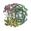
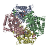
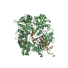
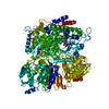
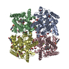
- PDB-6ayv: Crystal structure of fructose-1,6-bisphosphatase T84A from Mycoba... -
+ Open data

- Basic information
| Entry | Database: PDB / ID: 6ayv | ||||||
|---|---|---|---|---|---|---|---|
| Title | Crystal structure of fructose-1,6-bisphosphatase T84A from Mycobacterium tuberculosis | ||||||
 Components Components | Fructose-1,6-bisphosphatase class 2 | ||||||
 Keywords Keywords | HYDROLASE / Fructose-1 / 6-bisphosphatase / gluconeogenesis / Class II phosphatase / Mycobacterium tuberculosis | ||||||
| Function / homology |  Function and homology information Function and homology informationglycerol metabolic process / fructose-bisphosphatase / fructose 1,6-bisphosphate 1-phosphatase activity / fructose 1,6-bisphosphate metabolic process / gluconeogenesis / metal ion binding / cytosol Similarity search - Function | ||||||
| Biological species |   Mycobacterium tuberculosis (bacteria) Mycobacterium tuberculosis (bacteria) | ||||||
| Method |  X-RAY DIFFRACTION / SYNCHROTRON / MOLECULAR REPLACEMENT / Resolution: 2.3 Å X-RAY DIFFRACTION / SYNCHROTRON / MOLECULAR REPLACEMENT / Resolution: 2.3 Å | ||||||
 Authors Authors | Abad-Zapatero, C. / Wolf, N. / Gutka, H.J. / Movahedzadeh, F. | ||||||
| Funding support |  United States, 1items United States, 1items
| ||||||
 Citation Citation | Journal: Acta Crystallogr D Struct Biol / Year: 2018 Title: Structures of the Mycobacterium tuberculosis GlpX protein (class II fructose-1,6-bisphosphatase): implications for the active oligomeric state, catalytic mechanism and citrate inhibition. Authors: Wolf, N.M. / Gutka, H.J. / Movahedzadeh, F. / Abad-Zapatero, C. | ||||||
| History |
|
- Structure visualization
| Structure viewer | Molecule: MolmilJmol/JSmol |
|---|
- Downloads & links
-Download
| PDBx/mmCIF format | 6ayv.cif.gz | 142.1 KB | Display | PDBx/mmCIF format |
|---|---|---|---|---|
| PDB format | pdb6ayv.ent.gz | 109.6 KB | Display | PDB format |
| PDBx/mmJSON format | 6ayv.json.gz | Tree view | PDBx/mmJSON format | |
| Others |  Other downloads Other downloads |
-Validation report
| Arichive directory | https://data.pdbj.org/pub/pdb/validation_reports/ay/6ayvftp://data.pdbj.org/pub/pdb/validation_reports/ay/6ayv | HTTPS FTP |
|---|
-Related structure data
| Related structure data | 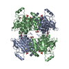 6ayuC 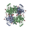 6ayyC 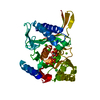 3d1rS S: Starting model for refinement C: citing same article ( |
|---|---|
| Similar structure data |


-Links
 PDBj
PDBj
- Assembly
| Deposited unit | 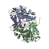
| ||||||||
|---|---|---|---|---|---|---|---|---|---|
| 1 | 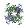
| ||||||||
| Unit cell |
|
-Components
-Protein / Sugars , 2 types, 4 molecules AB
| #1: Protein | Mass: 36630.406 Da / Num. of mol.: 2 / Fragment: UNP residues 35-362 / Mutation: T84A Source method: isolated from a genetically manipulated source Source: (gene. exp.) Mycobacterium tuberculosis (bacteria) / Strain: CDC 1551 / Oshkosh / Gene: glpX, MT1131 / Plasmid: pET15b / Cell (production host): BL21 (DE3) / Production host: #2: Sugar |  Type: D-saccharide, beta linking / Mass: 260.136 Da / Num. of mol.: 2 Type: D-saccharide, beta linking / Mass: 260.136 Da / Num. of mol.: 2Source method: isolated from a genetically manipulated source Formula: C6H13O9P |
|---|
-Non-polymers , 4 types, 318 molecules 


| #3: Chemical |  Mass: 24.305 Da / Num. of mol.: 3 / Source method: obtained synthetically / Formula: Mg Mass: 24.305 Da / Num. of mol.: 3 / Source method: obtained synthetically / Formula: Mg#4: Chemical | ChemComp-GOL /  Mass: 92.094 Da / Num. of mol.: 11 / Source method: obtained synthetically / Formula: C3H8O3 Mass: 92.094 Da / Num. of mol.: 11 / Source method: obtained synthetically / Formula: C3H8O3#5: Chemical | ChemComp-MLI / |  Mass: 102.046 Da / Num. of mol.: 1 / Source method: obtained synthetically / Formula: C3H2O4 Mass: 102.046 Da / Num. of mol.: 1 / Source method: obtained synthetically / Formula: C3H2O4#6: Water | ChemComp-HOH / | Mass: 18.015 Da / Num. of mol.: 303 / Source method: isolated from a natural source / Formula: H2O |
|---|
-Experimental details
-Experiment
| Experiment | Method: X-RAY DIFFRACTION / Number of used crystals: 1 |
|---|
- Sample preparation
| Crystal | Density Matthews: 2.45 Å3/Da / Density % sol: 49.71 % |
|---|---|
| Crystal grow | Temperature: 298 K / Method: vapor diffusion, hanging drop / pH: 6 / Details: 2.4 M sodium malonate pH 6.0 |
-Data collection
| Diffraction | Mean temperature: 100 K |
|---|---|
| Diffraction source | Source: SYNCHROTRON / Site: APS / Beamline: 21-ID-G / Wavelength: 0.97856 Å |
| Detector | Type: MARRESEARCH / Detector: CCD / Date: Dec 10, 2016 |
| Radiation | Protocol: SINGLE WAVELENGTH / Monochromatic (M) / Laue (L): M / Scattering type: x-ray |
| Radiation wavelength | Wavelength: 0.97856 Å / Relative weight: 1 |
| Reflection | Resolution: 2.3→40 Å / Num. obs: 33376 / % possible obs: 100 % / Redundancy: 23.7 % / Rmerge(I) obs: 0.125 / Rpim(I) all: 0.026 / Rsym value: 0.125 / Net I/σ(I): 55 |
| Reflection shell | Resolution: 2.3→2.34 Å / Redundancy: 21.4 % / Rmerge(I) obs: 2.314 / Mean I/σ(I) obs: 2.1 / Num. unique obs: 1613 / CC1/2: 0.691 / Rpim(I) all: 0.517 / Rsym value: 2.314 / Χ2: 0.95 / % possible all: 100 |
- Processing
| Software |
| |||||||||||||||||||||||||||||||||||||||||||||||||||||||||||||||||||||||||||||||||||||||||||
|---|---|---|---|---|---|---|---|---|---|---|---|---|---|---|---|---|---|---|---|---|---|---|---|---|---|---|---|---|---|---|---|---|---|---|---|---|---|---|---|---|---|---|---|---|---|---|---|---|---|---|---|---|---|---|---|---|---|---|---|---|---|---|---|---|---|---|---|---|---|---|---|---|---|---|---|---|---|---|---|---|---|---|---|---|---|---|---|---|---|---|---|---|
| Refinement | Method to determine structure: MOLECULAR REPLACEMENT Starting model: 3d1r Resolution: 2.3→38.768 Å / SU ML: 0.27 / Cross valid method: FREE R-VALUE / σ(F): 1.34 / Phase error: 26.45 / Stereochemistry target values: ML
| |||||||||||||||||||||||||||||||||||||||||||||||||||||||||||||||||||||||||||||||||||||||||||
| Solvent computation | Shrinkage radii: 0.9 Å / VDW probe radii: 1.11 Å / Solvent model: FLAT BULK SOLVENT MODEL | |||||||||||||||||||||||||||||||||||||||||||||||||||||||||||||||||||||||||||||||||||||||||||
| Refinement step | Cycle: LAST / Resolution: 2.3→38.768 Å
| |||||||||||||||||||||||||||||||||||||||||||||||||||||||||||||||||||||||||||||||||||||||||||
| Refine LS restraints |
| |||||||||||||||||||||||||||||||||||||||||||||||||||||||||||||||||||||||||||||||||||||||||||
| LS refinement shell |
|
