 Movie
Movie Controller

[English] 日本語
 Yorodumi
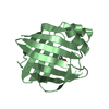
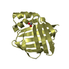
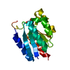
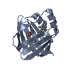
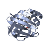
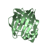
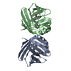
- PDB-6at8: 1.1 Angstrom Resolution Structure of Human Cellular Retinol-Bindi... -
+ Open data

- Basic information
| Entry | Database: PDB / ID: 6at8 | |||||||||
|---|---|---|---|---|---|---|---|---|---|---|
| Title | 1.1 Angstrom Resolution Structure of Human Cellular Retinol-Binding Protein IV | |||||||||
 Components Components | Retinoid-binding protein 7 | |||||||||
 Keywords Keywords | LIPID BINDING PROTEIN / Retinol / Lipid transport | |||||||||
| Function / homology |  Function and homology information Function and homology informationretinal binding / retinol binding / fatty acid transport / fatty acid binding / nucleus / cytosol Similarity search - Function | |||||||||
| Biological species |  Homo sapiens (human) Homo sapiens (human) | |||||||||
| Method |  X-RAY DIFFRACTION / SYNCHROTRON / MOLECULAR REPLACEMENT / Resolution: 1.105 Å X-RAY DIFFRACTION / SYNCHROTRON / MOLECULAR REPLACEMENT / Resolution: 1.105 Å | |||||||||
 Authors Authors | Silvaroli, J.A. / Pleshinger, M.J. / Kiser, P.D. / Golczak, M. | |||||||||
| Funding support |  United States, 1items United States, 1items
| |||||||||
 Citation Citation | Journal: To Be Published Title: 1.1 Angstrom Resolution Structure of Human Cellular Retinol-Binding Protein IV Authors: Silvaroli, J.A. / Pleshinger, M.J. / Kiser, P.D. / Golczak, M. | |||||||||
| History |
|
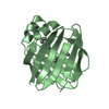
- Structure visualization
| Structure viewer | Molecule: MolmilJmol/JSmol |
|---|
- Downloads & links
-Download
| PDBx/mmCIF format | 6at8.cif.gz | 111.6 KB | Display | PDBx/mmCIF format |
|---|---|---|---|---|
| PDB format | pdb6at8.ent.gz | 86.6 KB | Display | PDB format |
| PDBx/mmJSON format | 6at8.json.gz | Tree view | PDBx/mmJSON format | |
| Others |  Other downloads Other downloads |
-Validation report
| Arichive directory | https://data.pdbj.org/pub/pdb/validation_reports/at/6at8ftp://data.pdbj.org/pub/pdb/validation_reports/at/6at8 | HTTPS FTP |
|---|
-Related structure data
| Related structure data | 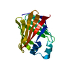 1lpjS S: Starting model for refinement |
|---|---|
| Similar structure data |


-Links
 PDBj
PDBj

- Assembly
| Deposited unit | 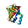
| |||||||||||||||
|---|---|---|---|---|---|---|---|---|---|---|---|---|---|---|---|---|
| 1 |
| |||||||||||||||
| Unit cell |
| |||||||||||||||
| Components on special symmetry positions |
|
-Components
| #1: Protein | Mass: 15894.110 Da / Num. of mol.: 1 Source method: isolated from a genetically manipulated source Source: (gene. exp.) Homo sapiens (human) / Gene: RBP7 / Production host:  |
|---|---|
| #2: Chemical | ChemComp-PEG /   Mass: 106.120 Da / Num. of mol.: 1 / Source method: obtained synthetically / Formula: C4H10O3 Mass: 106.120 Da / Num. of mol.: 1 / Source method: obtained synthetically / Formula: C4H10O3 |
| #3: Chemical | ChemComp-GOL /   Mass: 92.094 Da / Num. of mol.: 1 / Source method: obtained synthetically / Formula: C3H8O3 Mass: 92.094 Da / Num. of mol.: 1 / Source method: obtained synthetically / Formula: C3H8O3 |
| #4: Water | ChemComp-HOH /  Mass: 18.015 Da / Num. of mol.: 231 / Source method: isolated from a natural source / Formula: H2O Mass: 18.015 Da / Num. of mol.: 231 / Source method: isolated from a natural source / Formula: H2O |
-Experimental details
-Experiment
| Experiment | Method: X-RAY DIFFRACTION / Number of used crystals: 1 |
|---|
- Sample preparation
| Crystal | Density Matthews: 2.08 Å3/Da / Density % sol: 40.96 % |
|---|---|
| Crystal grow | Temperature: 295 K / Method: vapor diffusion, sitting drop / pH: 6.5 / Details: 0.2 M NaCl, 0.1 M Bis-Tris/HCl, 25% PEG3350 (w/v) |
-Data collection
| Diffraction | Mean temperature: 80 K |
|---|---|
| Diffraction source | Source: SYNCHROTRON / Site: APS / Beamline: 24-ID-E / Wavelength: 0.97919 Å |
| Detector | Type: DECTRIS EIGER X 16M / Detector: PIXEL / Date: Aug 20, 2017 |
| Radiation | Protocol: SINGLE WAVELENGTH / Monochromatic (M) / Laue (L): M / Scattering type: x-ray |
| Radiation wavelength | Wavelength: 0.97919 Å / Relative weight: 1 |
| Reflection | Resolution: 1.105→29.19 Å / Num. obs: 49226 / % possible obs: 94.4 % / Redundancy: 5.5 % / CC1/2: 0.994 / Rmerge(I) obs: 0.053 / Rpim(I) all: 0.026 / Rrim(I) all: 0.035 / Rsym value: 0.064 / Net I/σ(I): 31.8 |
| Reflection shell | Resolution: 1.105→1.16 Å / Mean I/σ(I) obs: 8.6 / Num. unique obs: 5474 / CC1/2: 0.982 / Rpim(I) all: 0.077 / Rrim(I) all: 0.09 / % possible all: 72 |
- Processing
| Software |
| ||||||||||||||||||||||||||||||||||||||||||||||||||||||||||||||||||||||||||||||||||||||||||||||||||||||||||||||||||||||||||||||
|---|---|---|---|---|---|---|---|---|---|---|---|---|---|---|---|---|---|---|---|---|---|---|---|---|---|---|---|---|---|---|---|---|---|---|---|---|---|---|---|---|---|---|---|---|---|---|---|---|---|---|---|---|---|---|---|---|---|---|---|---|---|---|---|---|---|---|---|---|---|---|---|---|---|---|---|---|---|---|---|---|---|---|---|---|---|---|---|---|---|---|---|---|---|---|---|---|---|---|---|---|---|---|---|---|---|---|---|---|---|---|---|---|---|---|---|---|---|---|---|---|---|---|---|---|---|---|---|
| Refinement | Method to determine structure: MOLECULAR REPLACEMENT Starting model: 1LPJ Resolution: 1.105→29.19 Å / SU ML: 0.07 / Cross valid method: FREE R-VALUE / σ(F): 1.36 / Phase error: 13.12
| ||||||||||||||||||||||||||||||||||||||||||||||||||||||||||||||||||||||||||||||||||||||||||||||||||||||||||||||||||||||||||||||
| Solvent computation | Shrinkage radii: 0.9 Å / VDW probe radii: 1.11 Å | ||||||||||||||||||||||||||||||||||||||||||||||||||||||||||||||||||||||||||||||||||||||||||||||||||||||||||||||||||||||||||||||
| Refinement step | Cycle: LAST / Resolution: 1.105→29.19 Å
| ||||||||||||||||||||||||||||||||||||||||||||||||||||||||||||||||||||||||||||||||||||||||||||||||||||||||||||||||||||||||||||||
| Refine LS restraints |
| ||||||||||||||||||||||||||||||||||||||||||||||||||||||||||||||||||||||||||||||||||||||||||||||||||||||||||||||||||||||||||||||
| LS refinement shell |
|
