 Movie
Movie Controller


+ Open data

- Basic information
| Entry | Database: PDB / ID: 6at4 | |||||||||
|---|---|---|---|---|---|---|---|---|---|---|
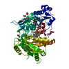
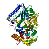
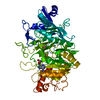
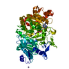
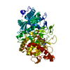
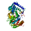
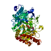
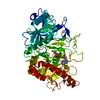
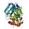
| Title | E. coli phosphoenolpyruvate carboxykinase bound to thiosulfate | |||||||||
 Components Components | Phosphoenolpyruvate carboxykinase (ATP) | |||||||||
 Keywords Keywords | LYASE / Nonnative ligand | |||||||||
| Function / homology |  Function and homology information Function and homology informationphosphoenolpyruvate carboxykinase (ATP) / phosphoenolpyruvate carboxykinase (ATP) activity / gluconeogenesis / calcium ion binding / magnesium ion binding / ATP binding / cytosol Similarity search - Function | |||||||||
| Biological species |  | |||||||||
| Method |  X-RAY DIFFRACTION / SYNCHROTRON / MOLECULAR REPLACEMENT / Resolution: 1.332 Å X-RAY DIFFRACTION / SYNCHROTRON / MOLECULAR REPLACEMENT / Resolution: 1.332 Å | |||||||||
 Authors Authors | Tang, H.Y.H. / Shin, D.S. / Tainer, J.A. | |||||||||
| Funding support |  United States, 2items United States, 2items
| |||||||||
 Citation Citation | Journal: Biochemistry / Year: 2018 Title: Structural Control of Nonnative Ligand Binding in Engineered Mutants of Phosphoenolpyruvate Carboxykinase. Authors: Tang, H.Y.H. / Shin, D.S. / Hura, G.L. / Yang, Y. / Hu, X. / Lightstone, F.C. / McGee, M.D. / Padgett, H.S. / Yannone, S.M. / Tainer, J.A. | |||||||||
| History |
|
- Structure visualization
| Structure viewer | Molecule: MolmilJmol/JSmol |
|---|
- Downloads & links
-Download
| PDBx/mmCIF format | 6at4.cif.gz | 600.6 KB | Display | PDBx/mmCIF format |
|---|---|---|---|---|
| PDB format | pdb6at4.ent.gz | 502.9 KB | Display | PDB format |
| PDBx/mmJSON format | 6at4.json.gz | Tree view | PDBx/mmJSON format | |
| Others |  Other downloads Other downloads |
-Validation report
| Summary document | 6at4_validation.pdf.gz | 2.1 MB | Display | wwPDB validaton report |
|---|---|---|---|---|
| Full document | 6at4_full_validation.pdf.gz | 2.1 MB | Display | |
| Data in XML | 6at4_validation.xml.gz | 45.3 KB | Display | |
| Data in CIF | 6at4_validation.cif.gz | 69.3 KB | Display | |
| Arichive directory | https://data.pdbj.org/pub/pdb/validation_reports/at/6at4ftp://data.pdbj.org/pub/pdb/validation_reports/at/6at4 | HTTPS FTP |
-Related structure data
| Related structure data |  6asiC  6asmC 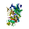 6asnC 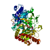 6at2C 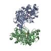 6at3C  2olqS S: Starting model for refinement C: citing same article ( |
|---|---|
| Similar structure data |

-Links
 PDBj
PDBj- Assembly
| Deposited unit | 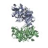
| ||||||||
|---|---|---|---|---|---|---|---|---|---|
| 1 | 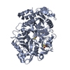
| ||||||||
| 2 | 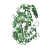
| ||||||||
| Unit cell |
|
-Components
| #1: Protein | Mass: 60538.066 Da / Num. of mol.: 2 Source method: isolated from a genetically manipulated source Source: (gene. exp.) References: UniProt: P22259, phosphoenolpyruvate carboxykinase (ATP) #2: Chemical | ChemComp-THJ /   Mass: 112.128 Da / Num. of mol.: 5 / Source method: obtained synthetically / Formula: O3S2 / Feature type: SUBJECT OF INVESTIGATION Mass: 112.128 Da / Num. of mol.: 5 / Source method: obtained synthetically / Formula: O3S2 / Feature type: SUBJECT OF INVESTIGATION#3: Water | ChemComp-HOH / |  Mass: 18.015 Da / Num. of mol.: 926 / Source method: isolated from a natural source / Formula: H2O Mass: 18.015 Da / Num. of mol.: 926 / Source method: isolated from a natural source / Formula: H2O |
|---|
-Experimental details
-Experiment
| Experiment | Method: X-RAY DIFFRACTION / Number of used crystals: 1 |
|---|
- Sample preparation
| Crystal | Density Matthews: 2.41 Å3/Da / Density % sol: 49.06 % |
|---|---|
| Crystal grow | Temperature: 293 K / Method: vapor diffusion, hanging drop Details: 20% PEG 3350, 0.4 M sodium chloride, 0.1 M Bis-Tris pH 5.5, 0.1 M sodium thiosulfate |
-Data collection
| Diffraction | Mean temperature: 100 K |
|---|---|
| Diffraction source | Source: SYNCHROTRON / Site: ALS / Beamline: 8.3.1 / Wavelength: 1.1158 Å |
| Detector | Type: DECTRIS PILATUS3 6M / Detector: PIXEL / Date: Jul 26, 2016 |
| Radiation | Protocol: SINGLE WAVELENGTH / Monochromatic (M) / Laue (L): M / Scattering type: x-ray |
| Radiation wavelength | Wavelength: 1.1158 Å / Relative weight: 1 |
| Reflection | Resolution: 1.332→119.156 Å / Num. obs: 470294 / % possible obs: 90.7 % / Redundancy: 3.5 % / CC1/2: 0.999 / Rmerge(I) obs: 0.047 / Net I/σ(I): 13.78 |
| Reflection shell | Resolution: 1.332→1.4 Å / Redundancy: 2.21 % / Rmerge(I) obs: 0.795 / Mean I/σ(I) obs: 1 / Num. unique obs: 48895 / CC1/2: 0.436 / % possible all: 58.2 |
- Processing
| Software |
| ||||||||||||||||||||||||||||||||||||||||||||||||||||||||||||||||||||||||||||||||||||||||||||||||||||||||||||||||||||||||||||||||||||||||||||||||||||||||||||||||||||||||||||||||||||||||||||||||||||
|---|---|---|---|---|---|---|---|---|---|---|---|---|---|---|---|---|---|---|---|---|---|---|---|---|---|---|---|---|---|---|---|---|---|---|---|---|---|---|---|---|---|---|---|---|---|---|---|---|---|---|---|---|---|---|---|---|---|---|---|---|---|---|---|---|---|---|---|---|---|---|---|---|---|---|---|---|---|---|---|---|---|---|---|---|---|---|---|---|---|---|---|---|---|---|---|---|---|---|---|---|---|---|---|---|---|---|---|---|---|---|---|---|---|---|---|---|---|---|---|---|---|---|---|---|---|---|---|---|---|---|---|---|---|---|---|---|---|---|---|---|---|---|---|---|---|---|---|---|---|---|---|---|---|---|---|---|---|---|---|---|---|---|---|---|---|---|---|---|---|---|---|---|---|---|---|---|---|---|---|---|---|---|---|---|---|---|---|---|---|---|---|---|---|---|---|---|---|
| Refinement | Method to determine structure: MOLECULAR REPLACEMENT Starting model: 2OLQ Resolution: 1.332→78.188 Å / SU ML: 0.13 / Cross valid method: FREE R-VALUE / σ(F): 1.35 / Phase error: 15.39
| ||||||||||||||||||||||||||||||||||||||||||||||||||||||||||||||||||||||||||||||||||||||||||||||||||||||||||||||||||||||||||||||||||||||||||||||||||||||||||||||||||||||||||||||||||||||||||||||||||||
| Solvent computation | Shrinkage radii: 0.9 Å / VDW probe radii: 1.11 Å | ||||||||||||||||||||||||||||||||||||||||||||||||||||||||||||||||||||||||||||||||||||||||||||||||||||||||||||||||||||||||||||||||||||||||||||||||||||||||||||||||||||||||||||||||||||||||||||||||||||
| Refinement step | Cycle: LAST / Resolution: 1.332→78.188 Å
| ||||||||||||||||||||||||||||||||||||||||||||||||||||||||||||||||||||||||||||||||||||||||||||||||||||||||||||||||||||||||||||||||||||||||||||||||||||||||||||||||||||||||||||||||||||||||||||||||||||
| Refine LS restraints |
| ||||||||||||||||||||||||||||||||||||||||||||||||||||||||||||||||||||||||||||||||||||||||||||||||||||||||||||||||||||||||||||||||||||||||||||||||||||||||||||||||||||||||||||||||||||||||||||||||||||
| LS refinement shell |
|
