 Movie
Movie Controller


+ Open data

- Basic information
| Entry | Database: PDB / ID: 6as4 | |||||||||
|---|---|---|---|---|---|---|---|---|---|---|
| Title | Structure of a phage anti-CRISPR protein | |||||||||
 Components Components | NHis AcrE1 anti-crispr protein | |||||||||
 Keywords Keywords | VIRAL PROTEIN / phage protein | |||||||||
| Function / homology | Uncharacterized protein Function and homology information Function and homology information | |||||||||
| Biological species |  Pseudomonas phage JBD5 (virus) Pseudomonas phage JBD5 (virus) | |||||||||
| Method |  X-RAY DIFFRACTION / SYNCHROTRON / MOLECULAR REPLACEMENT / Resolution: 2 Å X-RAY DIFFRACTION / SYNCHROTRON / MOLECULAR REPLACEMENT / Resolution: 2 Å | |||||||||
 Authors Authors | Shah, M. / Calmettes, C. / Pawluk, A. / Mejdani, M. / Davidson, A.R. / Maxwell, K.L. / Moraes, T.F. | |||||||||
| Funding support |  Canada, 2items Canada, 2items
| |||||||||
 Citation Citation | Journal: MBio / Year: 2017 Title: Disabling a Type I-E CRISPR-Cas Nuclease with a Bacteriophage-Encoded Anti-CRISPR Protein. Authors: Pawluk, A. / Shah, M. / Mejdani, M. / Calmettes, C. / Moraes, T.F. / Davidson, A.R. / Maxwell, K.L. | |||||||||
| History |
|
- Structure visualization
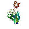
| Structure viewer | Molecule: MolmilJmol/JSmol |
|---|
- Downloads & links
-Download
| PDBx/mmCIF format | 6as4.cif.gz | 132 KB | Display | PDBx/mmCIF format |
|---|---|---|---|---|
| PDB format | pdb6as4.ent.gz | 103.4 KB | Display | PDB format |
| PDBx/mmJSON format | 6as4.json.gz | Tree view | PDBx/mmJSON format | |
| Others |  Other downloads Other downloads |
-Validation report
| Arichive directory | https://data.pdbj.org/pub/pdb/validation_reports/as/6as4ftp://data.pdbj.org/pub/pdb/validation_reports/as/6as4 | HTTPS FTP |
|---|
-Related structure data


-Links
 PDBj
PDBj- Assembly
| Deposited unit | 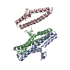
| ||||||||
|---|---|---|---|---|---|---|---|---|---|
| 1 | 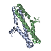
| ||||||||
| 2 | 
| ||||||||
| Unit cell |
|
-Components
| #1: Protein | Mass: 12121.561 Da / Num. of mol.: 3 Source method: isolated from a genetically manipulated source Details: Phage / Source: (gene. exp.) Pseudomonas phage JBD5 (virus) / Gene: JBD5_034 / Plasmid: pET21d(+) / Production host:  #2: Water | ChemComp-HOH / |  Mass: 18.015 Da / Num. of mol.: 150 / Source method: isolated from a natural source / Formula: H2O Mass: 18.015 Da / Num. of mol.: 150 / Source method: isolated from a natural source / Formula: H2O |
|---|
-Experimental details
-Experiment
| Experiment | Method: X-RAY DIFFRACTION / Number of used crystals: 1 |
|---|
- Sample preparation
| Crystal | Density Matthews: 2.47 Å3/Da / Density % sol: 50.23 % |
|---|---|
| Crystal grow | Temperature: 293 K / Method: vapor diffusion, sitting drop / pH: 7.5 / Details: 0.2M ammonium citrate dibasic, 20% PEG3350 |
-Data collection
| Diffraction | Mean temperature: 105 K |
|---|---|
| Diffraction source | Source: SYNCHROTRON / Site: CLSI / Beamline: 08B1-1 / Wavelength: 0.9793 Å |
| Detector | Type: RAYONIX MX300HE / Detector: CCD / Date: Jun 10, 2015 |
| Radiation | Protocol: SINGLE WAVELENGTH / Monochromatic (M) / Laue (L): M / Scattering type: x-ray |
| Radiation wavelength | Wavelength: 0.9793 Å / Relative weight: 1 |
| Reflection | Resolution: 2→47.585 Å / Num. obs: 23742 / % possible obs: 98.5 % / Redundancy: 7.7 % / Biso Wilson estimate: 37.05 Å2 / Rmerge(I) obs: 0.1033 / Net I/σ(I): 11.2 |
| Reflection shell | Resolution: 2→2.071 Å / Redundancy: 7.7 % / Rmerge(I) obs: 1.596 / Mean I/σ(I) obs: 1.57 / CC1/2: 0.529 / % possible all: 97.59 |
- Processing
| Software |
| ||||||||||||||||||||||||||||||||||||||||||||||||||||||||||||||||||||||||||||||||||||||||||||||||||||
|---|---|---|---|---|---|---|---|---|---|---|---|---|---|---|---|---|---|---|---|---|---|---|---|---|---|---|---|---|---|---|---|---|---|---|---|---|---|---|---|---|---|---|---|---|---|---|---|---|---|---|---|---|---|---|---|---|---|---|---|---|---|---|---|---|---|---|---|---|---|---|---|---|---|---|---|---|---|---|---|---|---|---|---|---|---|---|---|---|---|---|---|---|---|---|---|---|---|---|---|---|---|
| Refinement | Method to determine structure: MOLECULAR REPLACEMENT Starting model: AcrE1_CHis Resolution: 2→47.585 Å / SU ML: 0.24 / Cross valid method: FREE R-VALUE / σ(F): 1.35 / Phase error: 24.65
| ||||||||||||||||||||||||||||||||||||||||||||||||||||||||||||||||||||||||||||||||||||||||||||||||||||
| Solvent computation | Shrinkage radii: 0.9 Å / VDW probe radii: 1.11 Å | ||||||||||||||||||||||||||||||||||||||||||||||||||||||||||||||||||||||||||||||||||||||||||||||||||||
| Displacement parameters | Biso max: 144.89 Å2 / Biso mean: 48.1355 Å2 / Biso min: 22.15 Å2 | ||||||||||||||||||||||||||||||||||||||||||||||||||||||||||||||||||||||||||||||||||||||||||||||||||||
| Refinement step | Cycle: final / Resolution: 2→47.585 Å
| ||||||||||||||||||||||||||||||||||||||||||||||||||||||||||||||||||||||||||||||||||||||||||||||||||||
| Refine LS restraints |
| ||||||||||||||||||||||||||||||||||||||||||||||||||||||||||||||||||||||||||||||||||||||||||||||||||||
| LS refinement shell | Refine-ID: X-RAY DIFFRACTION / Rfactor Rfree error: 0 / Total num. of bins used: 8
| ||||||||||||||||||||||||||||||||||||||||||||||||||||||||||||||||||||||||||||||||||||||||||||||||||||
| Refinement TLS params. | Method: refined / Refine-ID: X-RAY DIFFRACTION
| ||||||||||||||||||||||||||||||||||||||||||||||||||||||||||||||||||||||||||||||||||||||||||||||||||||
| Refinement TLS group |
|
