 Movie
Movie Controller


+ Open data

- Basic information
| Entry | Database: PDB / ID: 6apj | ||||||
|---|---|---|---|---|---|---|---|
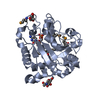
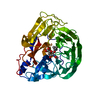
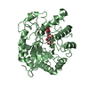
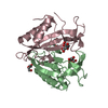
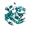
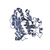
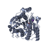
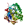
| Title | Crystal Structure of human ST6GALNAC2 | ||||||
 Components Components | Alpha-N-acetylgalactosaminide alpha-2,6-sialyltransferase 2 | ||||||
 Keywords Keywords | TRANSFERASE / glycosyltransferase / Structural Genomics / PSI-Biology / Northeast Structural Genomics Consortium / NESG | ||||||
| Function / homology |  Function and homology information Function and homology informationalpha-N-acetylgalactosaminide alpha-2,6-sialyltransferase / alpha-N-acetylgalactosaminide alpha-2,6-sialyltransferase activity / protein sialylation / Maturation of protein 3a / sialyltransferase activity / Maturation of protein 3a / protein O-linked glycosylation via N-acetylgalactosamine / Termination of O-glycan biosynthesis / Sialic acid metabolism / protein O-linked glycosylation ...alpha-N-acetylgalactosaminide alpha-2,6-sialyltransferase / alpha-N-acetylgalactosaminide alpha-2,6-sialyltransferase activity / protein sialylation / Maturation of protein 3a / sialyltransferase activity / Maturation of protein 3a / protein O-linked glycosylation via N-acetylgalactosamine / Termination of O-glycan biosynthesis / Sialic acid metabolism / protein O-linked glycosylation / glycoprotein biosynthetic process / Maturation of spike protein / viral protein processing / Golgi membrane Similarity search - Function | ||||||
| Biological species |  Homo sapiens (human) Homo sapiens (human) | ||||||
| Method |  X-RAY DIFFRACTION / SYNCHROTRON / SAD / Resolution: 3.1 Å X-RAY DIFFRACTION / SYNCHROTRON / SAD / Resolution: 3.1 Å | ||||||
 Authors Authors | Forouhar, F. / Moremen, K.W. / Northeast Structural Genomics Consortium (NESG) / Tong, L. | ||||||
 Citation Citation | Journal: Nat. Chem. Biol. / Year: 2018 Title: Expression system for structural and functional studies of human glycosylation enzymes. Authors: Moremen, K.W. / Ramiah, A. / Stuart, M. / Steel, J. / Meng, L. / Forouhar, F. / Moniz, H.A. / Gahlay, G. / Gao, Z. / Chapla, D. / Wang, S. / Yang, J.Y. / Prabhakar, P.K. / Johnson, R. / ...Authors: Moremen, K.W. / Ramiah, A. / Stuart, M. / Steel, J. / Meng, L. / Forouhar, F. / Moniz, H.A. / Gahlay, G. / Gao, Z. / Chapla, D. / Wang, S. / Yang, J.Y. / Prabhakar, P.K. / Johnson, R. / Rosa, M.D. / Geisler, C. / Nairn, A.V. / Seetharaman, J. / Wu, S.C. / Tong, L. / Gilbert, H.J. / LaBaer, J. / Jarvis, D.L. | ||||||
| History |
|
- Structure visualization
| Structure viewer | Molecule: MolmilJmol/JSmol |
|---|
- Downloads & links
-Download
| PDBx/mmCIF format | 6apj.cif.gz | 699.8 KB | Display | PDBx/mmCIF format |
|---|---|---|---|---|
| PDB format | pdb6apj.ent.gz | 582.9 KB | Display | PDB format |
| PDBx/mmJSON format | 6apj.json.gz | Tree view | PDBx/mmJSON format | |
| Others |  Other downloads Other downloads |
-Validation report
| Arichive directory | https://data.pdbj.org/pub/pdb/validation_reports/ap/6apjftp://data.pdbj.org/pub/pdb/validation_reports/ap/6apj | HTTPS FTP |
|---|
-Related structure data


-Links
 PDBj
PDBj- Assembly
| Deposited unit | 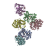
| ||||||||
|---|---|---|---|---|---|---|---|---|---|
| 1 | 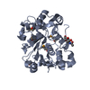
| ||||||||
| 2 | 
| ||||||||
| 3 | 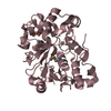
| ||||||||
| 4 | 
| ||||||||
| 5 | 
| ||||||||
| 6 | 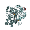
| ||||||||
| Unit cell |
|
-Components
| #1: Protein | Mass: 42224.582 Da / Num. of mol.: 6 Source method: isolated from a genetically manipulated source Source: (gene. exp.) Homo sapiens (human) / Gene: ST6GALNAC2, SIAT7B, SIATL1, STHM / Plasmid: pGEn2Details (production host): mammalian expression vector (CMV promoter) Cell line (production host): HEK293S GNT1- / Production host: Homo sapiens (human)References: UniProt: Q9UJ37, Transferases; Glycosyltransferases; Transferring other glycosyl groups #2: Sugar | ChemComp-NAG /   Type: D-saccharide, beta linking / Mass: 221.208 Da / Num. of mol.: 4 Type: D-saccharide, beta linking / Mass: 221.208 Da / Num. of mol.: 4Source method: isolated from a genetically manipulated source Formula: C8H15NO6 Has protein modification | Y | |
|---|
-Experimental details
-Experiment
| Experiment | Method: X-RAY DIFFRACTION / Number of used crystals: 1 |
|---|
- Sample preparation
| Crystal | Density Matthews: 2.52 Å3/Da / Density % sol: 51.28 % |
|---|---|
| Crystal grow | Temperature: 277 K / Method: microbatch / pH: 4.2 Details: 0.1M Sodium Citrate (pH 4.2), 0.1M Ammonium Sulfate, and 24% (w/v) PEG 20000 |
-Data collection
| Diffraction | Mean temperature: 100 K |
|---|---|
| Diffraction source | Source: SYNCHROTRON / Site: APS  / Beamline: 19-BM / Wavelength: 0.97921 Å / Beamline: 19-BM / Wavelength: 0.97921 Å |
| Detector | Type: ADSC QUANTUM 210r / Detector: CCD / Date: Mar 27, 2015 / Details: mirrors |
| Radiation | Monochromator: SI III / Protocol: SINGLE WAVELENGTH / Monochromatic (M) / Laue (L): M / Scattering type: x-ray |
| Radiation wavelength | Wavelength: 0.97921 Å / Relative weight: 1 |
| Reflection | Resolution: 3.1→128.4 Å / Num. obs: 52564 / % possible obs: 93.7 % / Observed criterion σ(F): 0 / Observed criterion σ(I): 0 / Redundancy: 3.6 % / Biso Wilson estimate: 91 Å2 / Rmerge(I) obs: 0.059 / Rpim(I) all: 0.059 / Rsym value: 0.083 / Χ2: 1.5 / Net I/av σ(I): 11.4 / Net I/σ(I): 11.4 |
| Reflection shell | Resolution: 3.1→3.24 Å / Redundancy: 3.7 % / Rmerge(I) obs: 0.588 / Mean I/σ(I) obs: 2.3 / Num. unique obs: 4117 / Rpim(I) all: 0.416 / Rsym value: 0.588 / Χ2: 0.9 / % possible all: 93.3 |
- Processing
| Software |
| |||||||||||||||||||||||||||||||||||||||||||||||||||||||||||||||||||||||||||||||||||||||||||||||||||||||||||||||||||||||||||||||||||||||||||||||||||||||||||||||||||||||||||||||||||||||||||||||||||||||||||||||||||||||||
|---|---|---|---|---|---|---|---|---|---|---|---|---|---|---|---|---|---|---|---|---|---|---|---|---|---|---|---|---|---|---|---|---|---|---|---|---|---|---|---|---|---|---|---|---|---|---|---|---|---|---|---|---|---|---|---|---|---|---|---|---|---|---|---|---|---|---|---|---|---|---|---|---|---|---|---|---|---|---|---|---|---|---|---|---|---|---|---|---|---|---|---|---|---|---|---|---|---|---|---|---|---|---|---|---|---|---|---|---|---|---|---|---|---|---|---|---|---|---|---|---|---|---|---|---|---|---|---|---|---|---|---|---|---|---|---|---|---|---|---|---|---|---|---|---|---|---|---|---|---|---|---|---|---|---|---|---|---|---|---|---|---|---|---|---|---|---|---|---|---|---|---|---|---|---|---|---|---|---|---|---|---|---|---|---|---|---|---|---|---|---|---|---|---|---|---|---|---|---|---|---|---|---|---|---|---|---|---|---|---|---|---|---|---|---|---|---|---|---|
| Refinement | Method to determine structure: SAD / Resolution: 3.1→44.28 Å / SU ML: 0.52 / Cross valid method: THROUGHOUT / σ(F): 2 / Phase error: 27.44
| |||||||||||||||||||||||||||||||||||||||||||||||||||||||||||||||||||||||||||||||||||||||||||||||||||||||||||||||||||||||||||||||||||||||||||||||||||||||||||||||||||||||||||||||||||||||||||||||||||||||||||||||||||||||||
| Solvent computation | Shrinkage radii: 0.9 Å / VDW probe radii: 1.11 Å | |||||||||||||||||||||||||||||||||||||||||||||||||||||||||||||||||||||||||||||||||||||||||||||||||||||||||||||||||||||||||||||||||||||||||||||||||||||||||||||||||||||||||||||||||||||||||||||||||||||||||||||||||||||||||
| Refinement step | Cycle: LAST / Resolution: 3.1→44.28 Å
| |||||||||||||||||||||||||||||||||||||||||||||||||||||||||||||||||||||||||||||||||||||||||||||||||||||||||||||||||||||||||||||||||||||||||||||||||||||||||||||||||||||||||||||||||||||||||||||||||||||||||||||||||||||||||
| Refine LS restraints |
| |||||||||||||||||||||||||||||||||||||||||||||||||||||||||||||||||||||||||||||||||||||||||||||||||||||||||||||||||||||||||||||||||||||||||||||||||||||||||||||||||||||||||||||||||||||||||||||||||||||||||||||||||||||||||
| LS refinement shell |
| |||||||||||||||||||||||||||||||||||||||||||||||||||||||||||||||||||||||||||||||||||||||||||||||||||||||||||||||||||||||||||||||||||||||||||||||||||||||||||||||||||||||||||||||||||||||||||||||||||||||||||||||||||||||||
| Refinement TLS params. | Method: refined / Origin x: -8.1163 Å / Origin y: 7.2187 Å / Origin z: 31.7724 Å
| |||||||||||||||||||||||||||||||||||||||||||||||||||||||||||||||||||||||||||||||||||||||||||||||||||||||||||||||||||||||||||||||||||||||||||||||||||||||||||||||||||||||||||||||||||||||||||||||||||||||||||||||||||||||||
| Refinement TLS group | Selection details: all |
