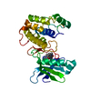
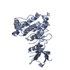
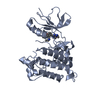
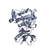
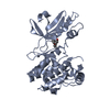
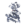
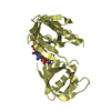
Entry Database : PDB / ID : 6ambTitle Crystal Structure of the Afadin RA1 domain in complex with HRAS Keywords / / / / Function / homology Function Domain/homology Component
/ / / / / / / / / / / / / / / / / / / / / / / / / / / / / / / / / / / / / / / / / / / / / / / / / / / / / / / / / / / / / / / / / / / / / / / / / / / / / / / / / / / / / / / / / / / / / / / / / / / / / / / / / / / / / / / / / / / / / / / / / / / / / / / / / / / / / / / / / / / / / / / / / / / / / Biological species Homo sapiens (human)Mus musculus (house mouse)Method / / / Resolution : 2.5 Å Authors Smith, M.J. / Ishiyama, N. / Ikura, M. Journal : Nat Commun / Year : 2017Title : Evolution of AF6-RAS association and its implications in mixed-lineage leukemia.Authors: Smith, M.J. / Ottoni, E. / Ishiyama, N. / Goudreault, M. / Haman, A. / Meyer, C. / Tucholska, M. / Gasmi-Seabrook, G. / Menezes, S. / Laister, R.C. / Minden, M.D. / Marschalek, R. / Gingras, ... Authors : Smith, M.J. / Ottoni, E. / Ishiyama, N. / Goudreault, M. / Haman, A. / Meyer, C. / Tucholska, M. / Gasmi-Seabrook, G. / Menezes, S. / Laister, R.C. / Minden, M.D. / Marschalek, R. / Gingras, A.C. / Hoang, T. / Ikura, M. History Deposition Aug 9, 2017 Deposition site / Processing site Revision 1.0 Nov 1, 2017 Provider / Type Revision 1.1 Nov 8, 2017 Group / Category / citation_authorItem _citation.journal_volume / _citation.page_first ... _citation.journal_volume / _citation.page_first / _citation.page_last / _citation.pdbx_database_id_PubMed / _citation.title / _citation_author.name Revision 1.2 Oct 4, 2023 Group / Database references / Refinement descriptionCategory chem_comp_atom / chem_comp_bond ... chem_comp_atom / chem_comp_bond / database_2 / pdbx_initial_refinement_model Item / _database_2.pdbx_database_accession
Show all Show less
 Movie
Movie Controller


 Open data
 Basic information
 Components
 Keywords
 Function and homology information
 Homo sapiens (human)
 X-RAY DIFFRACTION /
  Authors
 Citation
 Structure visualization
 Downloads & links
 Other downloads
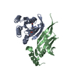


 PDBj
PDBj


 Assembly


 Mass: 522.196 Da / Num. of mol.: 1 / Source method: obtained synthetically / Formula: C10H17N6O13P3
 Mass: 24.305 Da / Num. of mol.: 1 / Source method: obtained synthetically / Formula: Mg
 Mass: 18.015 Da / Num. of mol.: 10 / Source method: isolated from a natural source / Formula: H2O
 Sample preparation
 / Beamline: 19-ID / Wavelength: 0.97929 Å
 Processing