 Movie
Movie Controller


+ Open data

- Basic information
| Entry | Database: PDB / ID: 6aip | ||||||
|---|---|---|---|---|---|---|---|
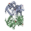
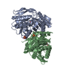
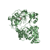
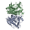
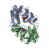
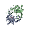
| Title | Cab2 mutant-H337A complex with phosphopantothenoylcystine | ||||||
 Components Components | Phosphopantothenate--cysteine ligase CAB2 | ||||||
 Keywords Keywords | LIGASE / Phosphopantothenate--cysteine ligase / complex / phosphopantothenoylcystine | ||||||
| Function / homology |  Function and homology information Function and homology informationCoA-synthesizing protein complex / Coenzyme A biosynthesis / phosphopantothenate-cysteine ligase (CTP) / phosphopantothenate--cysteine ligase activity / coenzyme A biosynthetic process / nucleus / cytoplasm Similarity search - Function | ||||||
| Biological species |  | ||||||
| Method |  X-RAY DIFFRACTION / SYNCHROTRON / MOLECULAR REPLACEMENT / Resolution: 1.99 Å X-RAY DIFFRACTION / SYNCHROTRON / MOLECULAR REPLACEMENT / Resolution: 1.99 Å | ||||||
 Authors Authors | Zheng, P. / Zhu, Z. | ||||||
 Citation Citation | Journal: J. Mol. Biol. / Year: 2019 Title: Crystallographic Analysis of the Catalytic Mechanism of Phosphopantothenoylcysteine Synthetase from Saccharomyces cerevisiae. Authors: Zheng, P. / Zhang, M. / Khan, M.H. / Liu, H. / Jin, Y. / Yue, J. / Gao, Y. / Teng, M. / Zhu, Z. / Niu, L. | ||||||
| History |
|
- Structure visualization
| Structure viewer | Molecule: MolmilJmol/JSmol |
|---|
- Downloads & links
-Download
| PDBx/mmCIF format | 6aip.cif.gz | 154 KB | Display | PDBx/mmCIF format |
|---|---|---|---|---|
| PDB format | pdb6aip.ent.gz | 118.1 KB | Display | PDB format |
| PDBx/mmJSON format | 6aip.json.gz | Tree view | PDBx/mmJSON format | |
| Others |  Other downloads Other downloads |
-Validation report
| Arichive directory | https://data.pdbj.org/pub/pdb/validation_reports/ai/6aipftp://data.pdbj.org/pub/pdb/validation_reports/ai/6aip | HTTPS FTP |
|---|
-Related structure data
| Related structure data |  6ai8C  6ai9C  6aikC  6aimC  1p9oS S: Starting model for refinement C: citing same article ( |
|---|---|
| Similar structure data |


-Links
 PDBj
PDBj- Assembly
| Deposited unit | 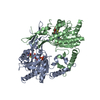
| ||||||||
|---|---|---|---|---|---|---|---|---|---|
| 1 |
| ||||||||
| Unit cell |
|
-Components
| #1: Protein | Mass: 42684.660 Da / Num. of mol.: 2 / Mutation: H337A Source method: isolated from a genetically manipulated source Source: (gene. exp.) Strain: ATCC 204508 / S288c / Gene: CAB2, YIL083C / Production host:  References: UniProt: P40506, phosphopantothenate-cysteine ligase (CTP) #2: Chemical |   Mass: 521.500 Da / Num. of mol.: 2 / Source method: obtained synthetically / Formula: C15H28N3O11PS2 / Feature type: SUBJECT OF INVESTIGATION Mass: 521.500 Da / Num. of mol.: 2 / Source method: obtained synthetically / Formula: C15H28N3O11PS2 / Feature type: SUBJECT OF INVESTIGATION#3: Water | ChemComp-HOH / |  Mass: 18.015 Da / Num. of mol.: 524 / Source method: isolated from a natural source / Formula: H2O Mass: 18.015 Da / Num. of mol.: 524 / Source method: isolated from a natural source / Formula: H2O |
|---|
-Experimental details
-Experiment
| Experiment | Method: X-RAY DIFFRACTION / Number of used crystals: 1 |
|---|
- Sample preparation
| Crystal | Density Matthews: 1.84 Å3/Da / Density % sol: 33.02 % |
|---|---|
| Crystal grow | Temperature: 287 K / Method: vapor diffusion, hanging drop / Details: 200mM ammonium tartrate, 20% PEG 3350 2mM MnCl2 |
-Data collection
| Diffraction | Mean temperature: 100 K |
|---|---|
| Diffraction source | Source: SYNCHROTRON / Site: SSRF  / Beamline: BL19U1 / Wavelength: 0.97853 Å / Beamline: BL19U1 / Wavelength: 0.97853 Å |
| Detector | Type: PSI PILATUS 6M / Detector: PIXEL / Date: Jan 26, 2018 |
| Radiation | Protocol: SINGLE WAVELENGTH / Monochromatic (M) / Laue (L): M / Scattering type: x-ray |
| Radiation wavelength | Wavelength: 0.97853 Å / Relative weight: 1 |
| Reflection | Resolution: 1.99→56.23 Å / Num. obs: 40420 / % possible obs: 96.2 % / Redundancy: 6.5 % / Rmerge(I) obs: 0.11 / Rpim(I) all: 0.0471 / Net I/σ(I): 16.7 |
| Reflection shell | Resolution: 2→2.03 Å / Rmerge(I) obs: 0.604 / Num. unique obs: 2030 / CC1/2: 0.873 / Rpim(I) all: 0.27 |
- Processing
| Software |
| ||||||||||||||||||||||||||||||||||||||||||||||||||||||||||||
|---|---|---|---|---|---|---|---|---|---|---|---|---|---|---|---|---|---|---|---|---|---|---|---|---|---|---|---|---|---|---|---|---|---|---|---|---|---|---|---|---|---|---|---|---|---|---|---|---|---|---|---|---|---|---|---|---|---|---|---|---|---|
| Refinement | Method to determine structure: MOLECULAR REPLACEMENT Starting model: 1P9O Resolution: 1.99→56.23 Å / Cor.coef. Fo:Fc: 0.956 / Cor.coef. Fo:Fc free: 0.94 / Cross valid method: THROUGHOUT / σ(F): 0 / ESU R: 0.221 / ESU R Free: 0.171 Details: HYDROGENS HAVE BEEN ADDED IN THE RIDING POSITIONS U VALUES : REFINED INDIVIDUALLY
| ||||||||||||||||||||||||||||||||||||||||||||||||||||||||||||
| Solvent computation | Ion probe radii: 0.8 Å / Shrinkage radii: 0.8 Å / VDW probe radii: 1.2 Å | ||||||||||||||||||||||||||||||||||||||||||||||||||||||||||||
| Displacement parameters | Biso max: 89.19 Å2 / Biso mean: 32.939 Å2 / Biso min: 17.42 Å2
| ||||||||||||||||||||||||||||||||||||||||||||||||||||||||||||
| Refinement step | Cycle: final / Resolution: 1.99→56.23 Å
| ||||||||||||||||||||||||||||||||||||||||||||||||||||||||||||
| Refine LS restraints |
| ||||||||||||||||||||||||||||||||||||||||||||||||||||||||||||
| LS refinement shell | Resolution: 1.989→2.04 Å / Rfactor Rfree error: 0 / Total num. of bins used: 20
|
