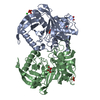
 Movie
Movie Controller


+ Open data

- Basic information
| Entry | Database: PDB / ID: 1p9o | ||||||
|---|---|---|---|---|---|---|---|
| Title | Crystal Structure of Phosphopantothenoylcysteine Synthetase | ||||||
 Components Components | Phosphopantothenoylcysteine synthetase | ||||||
 Keywords Keywords | LIGASE / synthetase | ||||||
| Function / homology |  Function and homology information Function and homology informationphosphopantothenate-cysteine ligase (ATP) / phosphopantothenate--cysteine ligase activity / Coenzyme A biosynthesis / acetyl-CoA biosynthetic process / heart process / coenzyme A biosynthetic process / protein homodimerization activity / ATP binding / identical protein binding / nucleus ...phosphopantothenate-cysteine ligase (ATP) / phosphopantothenate--cysteine ligase activity / Coenzyme A biosynthesis / acetyl-CoA biosynthetic process / heart process / coenzyme A biosynthetic process / protein homodimerization activity / ATP binding / identical protein binding / nucleus / cytoplasm / cytosol Similarity search - Function | ||||||
| Biological species |  Homo sapiens (human) Homo sapiens (human) | ||||||
| Method |  X-RAY DIFFRACTION / SYNCHROTRON / MOLECULAR REPLACEMENT / Resolution: 2.3 Å X-RAY DIFFRACTION / SYNCHROTRON / MOLECULAR REPLACEMENT / Resolution: 2.3 Å | ||||||
 Authors Authors | Manoj, N. / Strauss, E. / Begley, T.P. / Ealick, S.E. | ||||||
 Citation Citation | Journal: Structure / Year: 2003 Title: Structure of human phosphopantothenoylcysteine synthetase at 2.3 A resolution. Authors: Manoj, N. / Strauss, E. / Begley, T.P. / Ealick, S.E. | ||||||
| History |
| ||||||
| Remark 999 | SEQUENCE an appropriate sequence database reference for the protein was not available at the time ...SEQUENCE an appropriate sequence database reference for the protein was not available at the time of processing. |
- Structure visualization
| Structure viewer | Molecule: MolmilJmol/JSmol |
|---|
- Downloads & links
-Download
| PDBx/mmCIF format | 1p9o.cif.gz | 118.4 KB | Display | PDBx/mmCIF format |
|---|---|---|---|---|
| PDB format | pdb1p9o.ent.gz | 90.8 KB | Display | PDB format |
| PDBx/mmJSON format | 1p9o.json.gz | Tree view | PDBx/mmJSON format | |
| Others |  Other downloads Other downloads |
-Validation report
| Arichive directory | https://data.pdbj.org/pub/pdb/validation_reports/p9/1p9oftp://data.pdbj.org/pub/pdb/validation_reports/p9/1p9o | HTTPS FTP |
|---|
-Related structure data
| Similar structure data |
|---|


-Links
 PDBj
PDBj- Assembly
| Deposited unit | 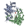
| ||||||||
|---|---|---|---|---|---|---|---|---|---|
| 1 |
| ||||||||
| Unit cell |
| ||||||||
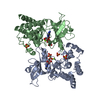
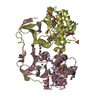
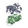
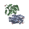
| Details | The crystallographic asymmetric unit contains two chains forming a homodimer that represents the biological unit |
-Components
| #1: Protein | Mass: 34164.176 Da / Num. of mol.: 2 Source method: isolated from a genetically manipulated source Source: (gene. exp.) Homo sapiens (human) / Gene: coaB / Plasmid: pPROEX-Hta / Production host:  References: UniProt: Q9HAB8, phosphopantothenate-cysteine ligase (CTP) #2: Chemical |   Mass: 96.063 Da / Num. of mol.: 2 / Source method: obtained synthetically / Formula: SO4 Mass: 96.063 Da / Num. of mol.: 2 / Source method: obtained synthetically / Formula: SO4#3: Water | ChemComp-HOH / |  Mass: 18.015 Da / Num. of mol.: 218 / Source method: isolated from a natural source / Formula: H2O Mass: 18.015 Da / Num. of mol.: 218 / Source method: isolated from a natural source / Formula: H2O |
|---|
-Experimental details
-Experiment
| Experiment | Method: X-RAY DIFFRACTION / Number of used crystals: 1 |
|---|
- Sample preparation
| Crystal | Density Matthews: 2.76 Å3/Da / Density % sol: 55.4 % | ||||||||||||||||||||||||||||||
|---|---|---|---|---|---|---|---|---|---|---|---|---|---|---|---|---|---|---|---|---|---|---|---|---|---|---|---|---|---|---|---|
| Crystal grow | Temperature: 298 K / Method: vapor diffusion, hanging drop / pH: 8.5 Details: PEG MME 2000, Ammonium Sulphate, Tris, pH 8.5, VAPOR DIFFUSION, HANGING DROP, temperature 298.0K | ||||||||||||||||||||||||||||||
| Crystal grow | *PLUS Method: vapor diffusion, hanging drop | ||||||||||||||||||||||||||||||
| Components of the solutions | *PLUS
|
-Data collection
| Diffraction | Mean temperature: 100 K |
|---|---|
| Diffraction source | Source: SYNCHROTRON / Site: APS  / Beamline: 8-BM / Wavelength: 0.9793 Å / Beamline: 8-BM / Wavelength: 0.9793 Å |
| Detector | Type: ADSC QUANTUM 315 / Detector: CCD / Date: Aug 5, 2002 |
| Radiation | Monochromator: SI (111) / Protocol: SINGLE WAVELENGTH / Monochromatic (M) / Laue (L): M / Scattering type: x-ray |
| Radiation wavelength | Wavelength: 0.9793 Å / Relative weight: 1 |
| Reflection | Resolution: 2.3→50 Å / Num. all: 35261 / Num. obs: 34732 / % possible obs: 98.5 % / Observed criterion σ(F): 0 / Observed criterion σ(I): 0 / Redundancy: 5.7 % / Biso Wilson estimate: 34 Å2 / Rsym value: 0.064 / Net I/σ(I): 31.2 |
| Reflection shell | Resolution: 2.3→2.38 Å / Redundancy: 5.6 % / Mean I/σ(I) obs: 6.3 / Num. unique all: 3418 / Rsym value: 0.313 / % possible all: 99.7 |
| Reflection | *PLUS Num. measured all: 199382 / Rmerge(I) obs: 0.064 |
| Reflection shell | *PLUS % possible obs: 99.7 % / Rmerge(I) obs: 0.313 |
- Processing
| Software |
| ||||||||||||||||||||||||||||||||||||
|---|---|---|---|---|---|---|---|---|---|---|---|---|---|---|---|---|---|---|---|---|---|---|---|---|---|---|---|---|---|---|---|---|---|---|---|---|---|
| Refinement | Method to determine structure: MOLECULAR REPLACEMENT Starting model: SAD phased structure of phosphopantothenoylcysteine synthetase Resolution: 2.3→45.32 Å / Rfactor Rfree error: 0.005 / Isotropic thermal model: RESTRAINED / Cross valid method: THROUGHOUT / σ(F): 0 / Stereochemistry target values: Engh & Huber
| ||||||||||||||||||||||||||||||||||||
| Solvent computation | Solvent model: FLAT MODEL / Bsol: 60.9858 Å2 / ksol: 0.35878 e/Å3 | ||||||||||||||||||||||||||||||||||||
| Displacement parameters | Biso mean: 43 Å2
| ||||||||||||||||||||||||||||||||||||
| Refine analyze |
| ||||||||||||||||||||||||||||||||||||
| Refinement step | Cycle: LAST / Resolution: 2.3→45.32 Å
| ||||||||||||||||||||||||||||||||||||
| Refine LS restraints |
| ||||||||||||||||||||||||||||||||||||
| LS refinement shell | Resolution: 2.3→2.44 Å / Rfactor Rfree error: 0.021 / Total num. of bins used: 6
| ||||||||||||||||||||||||||||||||||||
| Xplor file |
| ||||||||||||||||||||||||||||||||||||
| Refinement | *PLUS Highest resolution: 2.3 Å / Lowest resolution: 50 Å / % reflection Rfree: 7 % / Rfactor Rfree: 0.271 | ||||||||||||||||||||||||||||||||||||
| Solvent computation | *PLUS | ||||||||||||||||||||||||||||||||||||
| Displacement parameters | *PLUS | ||||||||||||||||||||||||||||||||||||
| Refine LS restraints | *PLUS
|
