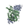
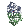
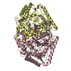
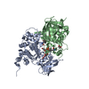
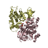
- PDB-6ae3: Crystal structure of GSK3beta complexed with Morin -
+
Open data
ID or keywords:
Loading...
-
Basic information
Entry
Database: PDB / ID: 6ae3
Title
Crystal structure of GSK3beta complexed with Morin
Components
Glycogen synthase kinase-3 beta
Keywords
SIGNALING PROTEIN / Kinase / Flavonoid
Function / homology
Function and homology information
hepatic stellate cell activation / negative regulation of synaptic assembly at neuromuscular junction / Regulation of HSF1-mediated heat shock response / negative regulation of protein localization to centrosome / B-WICH complex positively regulates rRNA expression / negative regulation of neuron maturation / Beta-catenin phosphorylation cascade / re-entry into mitotic cell cycle / CRMPs in Sema3A signaling / positive regulation of synaptic assembly at neuromuscular junction ...hepatic stellate cell activation / negative regulation of synaptic assembly at neuromuscular junction / Regulation of HSF1-mediated heat shock response / negative regulation of protein localization to centrosome / B-WICH complex positively regulates rRNA expression / negative regulation of neuron maturation / Beta-catenin phosphorylation cascade / re-entry into mitotic cell cycle / CRMPs in Sema3A signaling / positive regulation of synaptic assembly at neuromuscular junction / Disassembly of the destruction complex and recruitment of AXIN to the membrane / positive regulation of osteoclast proliferation / Transcriptional and post-translational regulation of MITF-M expression and activity / negative regulation of neuron migration / cell growth involved in cardiac muscle cell development / GSK3B and BTRC:CUL1-mediated-degradation of NFE2L2 / myotube differentiation / Degradation of beta-catenin by the destruction complex / protein localization to microtubule / neuron projection organization / regulation of microtubule anchoring at centrosome / negative regulation of mesenchymal stem cell differentiation / negative regulation of dendrite morphogenesis / negative regulation of dendrite development / superior temporal gyrus development / positive regulation of protein localization to cilium / response to methamphetamine hydrochloride / positive regulation of stem cell differentiation / negative regulation of glycogen biosynthetic process / negative regulation of TORC2 signaling / negative regulation of cardiac muscle hypertrophy / negative regulation of dopaminergic neuron differentiation / positive regulation of protein localization to centrosome / GLI3 is processed to GLI3R by the proteasome / maintenance of cell polarity / positive regulation of cardiac muscle cell differentiation / positive regulation of cilium assembly / tau-protein kinase / autosome genomic imprinting / beta-catenin destruction complex / bone remodeling / myoblast fusion / positive regulation of mitochondrial outer membrane permeabilization involved in apoptotic signaling pathway / regulation of protein export from nucleus / regulation of modification of postsynaptic structure / cellular response to interleukin-3 / positive regulation of osteoclast differentiation / phosphorylation / TORC2 signaling / meiosis I / regulation of long-term synaptic potentiation / Wnt signalosome / negative regulation of TOR signaling / meiotic spindle / regulation of microtubule-based process / regulation of axon extension / negative regulation of calcineurin-NFAT signaling cascade / positive regulation of mitochondrial membrane potential / cellular response to glucocorticoid stimulus / negative regulation of protein localization to nucleus / cellular response to hepatocyte growth factor stimulus / positive regulation of DNA biosynthetic process / negative regulation of epithelial to mesenchymal transition / tau-protein kinase activity / positive regulation of cell-matrix adhesion / response to zinc ion / glycogen metabolic process / ER overload response / cytoplasmic translational initiation / regulation of osteoblast differentiation / positive regulation of osteoblast proliferation / regulation of axonogenesis / regulation of dendrite morphogenesis / regulation of neuron projection development / hepatocyte apoptotic process / protein kinase A catalytic subunit binding / SCF-dependent proteasomal ubiquitin-dependent protein catabolic process / establishment of cell polarity / regulation of neurotransmitter receptor localization to postsynaptic specialization membrane / establishment or maintenance of cell polarity / negative regulation of smooth muscle cell apoptotic process / dynein complex binding / dynactin binding / intrinsic apoptotic signaling pathway in response to endoplasmic reticulum stress / epithelial to mesenchymal transition / canonical Wnt signaling pathway / regulation of neuronal synaptic plasticity / negative regulation of osteoblast differentiation / fat cell differentiation / NF-kappaB binding / negative regulation of ubiquitin-dependent protein catabolic process / negative regulation of extrinsic apoptotic signaling pathway via death domain receptors / conditioned place preference / positive regulation of axon extension / negative regulation of protein-containing complex assembly / extrinsic apoptotic signaling pathway / cellular response to retinoic acid / extrinsic apoptotic signaling pathway in absence of ligand / positive regulation of type I interferon production / cytoskeleton organization Similarity search - Function
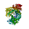
Glycogen synthase kinase 3, catalytic domain / : / Phosphorylase Kinase; domain 1 / Phosphorylase Kinase; domain 1 / Transferase(Phosphotransferase) domain 1 / Transferase(Phosphotransferase); domain 1 / Serine/threonine-protein kinase, active site / Serine/Threonine protein kinases active-site signature. / Protein kinase domain / Serine/Threonine protein kinases, catalytic domain ...Glycogen synthase kinase 3, catalytic domain / : / Phosphorylase Kinase; domain 1 / Phosphorylase Kinase; domain 1 / Transferase(Phosphotransferase) domain 1 / Transferase(Phosphotransferase); domain 1 / Serine/threonine-protein kinase, active site / Serine/Threonine protein kinases active-site signature. / Protein kinase domain / Serine/Threonine protein kinases, catalytic domain / Protein kinase, ATP binding site / Protein kinases ATP-binding region signature. / Protein kinase domain profile. / Protein kinase domain / Protein kinase-like domain superfamily / 2-Layer Sandwich / Orthogonal Bundle / Mainly Alpha / Alpha Beta Similarity search - Domain/homology
 Movie
Movie Controller


 Open data
 Basic information
 Components
 Keywords
 Function and homology information
 X-RAY DIFFRACTION /
  Authors
 Citation
 Structure visualization
 Downloads & links
 Other downloads


 PDBj
PDBj


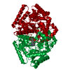
 Assembly

 Spodoptera frugiperda (fall armyworm)
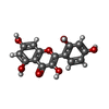
 Mass: 302.236 Da / Num. of mol.: 2 / Source method: obtained synthetically / Formula: C15H10O7 / Feature type: SUBJECT OF INVESTIGATION
 Mass: 92.094 Da / Num. of mol.: 4 / Source method: obtained synthetically / Formula: C3H8O3
 Mass: 18.015 Da / Num. of mol.: 422 / Source method: isolated from a natural source / Formula: H2O
 Sample preparation
 / Beamline: 4A / Wavelength: 0.987 Å
 Processing