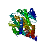
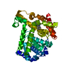
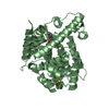
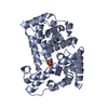
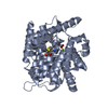
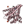
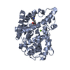
[English] 日本語
Yorodumi
- PDB-6acb: Crystal structure of PDE5 in complex with inhibitor LW1805 -

+
Open data


ID or keywords:

Loading...

-
Basic information

Entry
Database: PDB / ID: 6acb
TitleCrystal structure of PDE5 in complex with inhibitor LW1805
ComponentscGMP-specific 3',5'-cyclic phosphodiesterase
KeywordsHYDROLASE/HYDROLASE INHIBITOR / PDE5 in complex with inhibitor LW1805 / HYDROLASE-HYDROLASE INHIBITOR complex
Function / homology
Function and homology information


positive regulation of oocyte development / 3',5'-cyclic-GMP phosphodiesterase / regulation of nitric oxide mediated signal transduction / negative regulation of cardiac muscle contraction / oocyte development / RHOBTB1 GTPase cycle / relaxation of cardiac muscle / positive regulation of cardiac muscle hypertrophy / cGMP catabolic process / cGMP effects ...positive regulation of oocyte development / 3',5'-cyclic-GMP phosphodiesterase / regulation of nitric oxide mediated signal transduction / negative regulation of cardiac muscle contraction / oocyte development / RHOBTB1 GTPase cycle / relaxation of cardiac muscle / positive regulation of cardiac muscle hypertrophy / cGMP catabolic process / cGMP effects / 3',5'-cyclic-GMP phosphodiesterase activity / cGMP binding / 3',5'-cyclic-nucleotide phosphodiesterase activity / Smooth Muscle Contraction / T cell proliferation / negative regulation of T cell proliferation / signal transduction / metal ion binding / cytosol
Similarity search - Function
Catalytic domain of cyclic nucleotide phosphodiesterase 4b2b / 3'5'-cyclic nucleotide phosphodiesterase, catalytic domain / 3'5'-cyclic nucleotide phosphodiesterase / GAF domain / 3'5'-cyclic nucleotide phosphodiesterase, catalytic domain / 3'5'-cyclic nucleotide phosphodiesterase, conserved site / 3'5'-cyclic nucleotide phosphodiesterase, catalytic domain superfamily / 3'5'-cyclic nucleotide phosphodiesterase / 3'5'-cyclic nucleotide phosphodiesterase domain signature. / 3'5'-cyclic nucleotide phosphodiesterase domain profile. ...Catalytic domain of cyclic nucleotide phosphodiesterase 4b2b / 3'5'-cyclic nucleotide phosphodiesterase, catalytic domain / 3'5'-cyclic nucleotide phosphodiesterase / GAF domain / 3'5'-cyclic nucleotide phosphodiesterase, catalytic domain / 3'5'-cyclic nucleotide phosphodiesterase, conserved site / 3'5'-cyclic nucleotide phosphodiesterase, catalytic domain superfamily / 3'5'-cyclic nucleotide phosphodiesterase / 3'5'-cyclic nucleotide phosphodiesterase domain signature. / 3'5'-cyclic nucleotide phosphodiesterase domain profile. / Domain present in phytochromes and cGMP-specific phosphodiesterases. / GAF domain / GAF-like domain superfamily / Metal dependent phosphohydrolases with conserved 'HD' motif. / HD/PDEase domain / Orthogonal Bundle / Mainly Alpha
Similarity search - Domain/homology
Chem-9T9 / cGMP-specific 3',5'-cyclic phosphodiesterase
Similarity search - Component
Biological speciesHomo sapiens (human)
MethodX-RAY DIFFRACTION / MOLECULAR REPLACEMENT / Resolution: 2.8 Å
AuthorsWu, D. / Huang, Y.D. / Huang, Y.Y. / Luo, H.B.
Funding support China, 2items
OrganizationGrant numberCountry
National Natural Science Foundation of China81602955 China
National Natural Science Foundation of China21702238 China
CitationJournal: J. Med. Chem. / Year: 2018
Title: Optimization of Chromeno[2,3- c]pyrrol-9(2 H)-ones as Highly Potent, Selective, and Orally Bioavailable PDE5 Inhibitors: Structure-Activity Relationship, X-ray Crystal Structure, and ...Title: Optimization of Chromeno[2,3- c]pyrrol-9(2 H)-ones as Highly Potent, Selective, and Orally Bioavailable PDE5 Inhibitors: Structure-Activity Relationship, X-ray Crystal Structure, and Pharmacodynamic Effect on Pulmonary Arterial Hypertension.
Authors: Wu, D. / Huang, Y. / Chen, Y. / Huang, Y.Y. / Geng, H. / Zhang, T. / Zhang, C. / Li, Z. / Guo, L. / Chen, J. / Luo, H.B.
History
DepositionJul 26, 2018Deposition site: PDBJ / Processing site: PDBJ
Revision 1.0Sep 19, 2018Provider: repository / Type: Initial release
Revision 1.1Oct 10, 2018Group: Data collection / Database references / Structure summary
Category: citation / entity
Item: _citation.journal_volume / _citation.page_first ..._citation.journal_volume / _citation.page_first / _citation.page_last / _citation.title / _entity.formula_weight
Revision 1.2Nov 22, 2023Group: Data collection / Database references ...Data collection / Database references / Derived calculations / Refinement description / Structure summary
Category: chem_comp_atom / chem_comp_bond ...chem_comp_atom / chem_comp_bond / database_2 / entity / pdbx_initial_refinement_model / struct_conn
Item: _database_2.pdbx_DOI / _database_2.pdbx_database_accession ..._database_2.pdbx_DOI / _database_2.pdbx_database_accession / _entity.formula_weight / _struct_conn.pdbx_dist_value / _struct_conn.ptnr1_label_atom_id / _struct_conn.ptnr2_auth_comp_id / _struct_conn.ptnr2_auth_seq_id / _struct_conn.ptnr2_label_asym_id / _struct_conn.ptnr2_label_atom_id / _struct_conn.ptnr2_label_comp_id

-
Structure visualization

Structure viewerMolecule:
MolmilJmol/JSmol

Downloads & links

-
Assembly

Deposited unit
A: cGMP-specific 3',5'-cyclic phosphodiesterase
hetero molecules


Theoretical massNumber of molelcules
Total (without water)38,5365
Polymers37,7891
Non-polymers7474
Water81145
1


  • Idetical with deposited unit
  • defined by author&software
  • Evidence: cGMP-specific 3',5'-cyclic phosphodiesterase
TypeNameSymmetry operationNumber
identity operation1_555x,y,z1
Buried area330 Å2
ΔGint-54 kcal/mol
Surface area14410 Å2
MethodPISA
Unit cell
Length a, b, c (Å)74.577, 74.577, 132.092
Angle α, β, γ (deg.)90.00, 90.00, 120.00
Int Tables number152
Space group name H-MP3121

-
Components

-
Protein , 1 types, 1 molecules A

#1: Protein cGMP-specific 3',5'-cyclic phosphodiesterase / cGMP-binding cGMP-specific phosphodiesterase / CGB-PDE


Mass: 37788.531 Da / Num. of mol.: 1
Source method: isolated from a genetically manipulated source
Source: (gene. exp.) Homo sapiens (human) / Gene: PDE5A, PDE5 / Production host: Escherichia coli BL21 (bacteria) / Strain (production host): BL21
References: UniProt: O76074, 3',5'-cyclic-GMP phosphodiesterase

-
Non-polymers , 5 types, 49 molecules

#2: Chemical ChemComp-SO4 / SULFATE ION / Sulfate


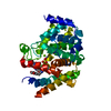
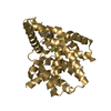
Mass: 96.063 Da / Num. of mol.: 1 / Source method: obtained synthetically / Formula: SO4
#3: Chemical ChemComp-ZN / ZINC ION


Mass: 65.409 Da / Num. of mol.: 1 / Source method: obtained synthetically / Formula: Zn
#4: Chemical ChemComp-MG / MAGNESIUM ION


Mass: 24.305 Da / Num. of mol.: 1 / Source method: obtained synthetically / Formula: Mg
#5: Chemical ChemComp-9T9 / 3-[(2H-1,3-benzodioxol-5-yl)methyl]-8-fluoro-6-{[2-(4-methylpiperazin-1-yl)ethyl]amino}-1-(1,3-thiazol-2-yl)[1]benzopyrano[2,3-c]pyrrol-9(2H)-one


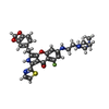
Mass: 561.627 Da / Num. of mol.: 1 / Source method: obtained synthetically / Formula: C29H28FN5O4S
#6: Water ChemComp-HOH / water / Water


Mass: 18.015 Da / Num. of mol.: 45 / Source method: isolated from a natural source / Formula: H2O

-
Experimental details

-
Experiment

ExperimentMethod: X-RAY DIFFRACTION / Number of used crystals: 1

-
Sample preparation

CrystalDensity Matthews: 2.81 Å3/Da / Density % sol: 56.17 %
Crystal growTemperature: 298 K / Method: vapor diffusion, hanging drop / pH: 6.5
Details: 0.1 M Cacodylate Sodium (pH6.5), 0.2 M MgSO4, 18% PEG3350, 2.5% ethanol

-
Data collection

DiffractionMean temperature: 100 K
Diffraction sourceSource: SEALED TUBE / Type: OXFORD DIFFRACTION NOVA / Wavelength: 1.5406 Å
DetectorType: OXFORD ONYX CCD / Detector: CCD / Date: Feb 10, 2018
RadiationProtocol: SINGLE WAVELENGTH / Monochromatic (M) / Laue (L): M / Scattering type: x-ray
Radiation wavelengthWavelength: 1.5406 Å / Relative weight: 1
ReflectionResolution: 2.8→24 Å / Num. obs: 10461 / % possible obs: 95.74 % / Redundancy: 7.9 % / Rmerge(I) obs: 0.132 / Net I/σ(I): 14.1
Reflection shellResolution: 2.8→2.9 Å

-
Processing

Software
NameVersionClassification
REFMAC5.8.0189refinement
CrysalisPro38.41data reduction
CrysalisPro38.41data scaling
PHASER1.1phasing
RefinementMethod to determine structure: MOLECULAR REPLACEMENT
Starting model: 4MD6
Resolution: 2.8→24 Å / Cor.coef. Fo:Fc: 0.929 / Cor.coef. Fo:Fc free: 0.867 / SU B: 16.778 / SU ML: 0.316 / Cross valid method: THROUGHOUT / ESU R Free: 0.397 / Details: HYDROGENS HAVE BEEN ADDED IN THE RIDING POSITIONS
RfactorNum. reflection% reflectionSelection details
Rfree0.26828 546 5.2 %RANDOM
Rwork0.1976 ---
obs0.20112 9913 95.52 %-
Solvent computationIon probe radii: 0.8 Å / Shrinkage radii: 0.8 Å / VDW probe radii: 1.2 Å
Displacement parametersBiso mean: 41.812 Å2
Baniso -1Baniso -2Baniso -3
1--0.02 Å2-0.01 Å2-0 Å2
2---0.02 Å20 Å2
3---0.06 Å2
Refinement stepCycle: 1 / Resolution: 2.8→24 Å
ProteinNucleic acidLigandSolventTotal
Num. atoms2499 0 47 45 2591
Refine LS restraints
Refine-IDTypeDev idealDev ideal targetNumber
X-RAY DIFFRACTIONr_bond_refined_d0.0110.0192598
X-RAY DIFFRACTIONr_bond_other_d0.0020.022406
X-RAY DIFFRACTIONr_angle_refined_deg1.511.9433514
X-RAY DIFFRACTIONr_angle_other_deg1.00135591
X-RAY DIFFRACTIONr_dihedral_angle_1_deg7.9885306
X-RAY DIFFRACTIONr_dihedral_angle_2_deg42.59324.8125
X-RAY DIFFRACTIONr_dihedral_angle_3_deg17.53515474
X-RAY DIFFRACTIONr_dihedral_angle_4_deg14.8371513
X-RAY DIFFRACTIONr_chiral_restr0.0840.2390
X-RAY DIFFRACTIONr_gen_planes_refined0.0060.022970
X-RAY DIFFRACTIONr_gen_planes_other0.0020.02515
X-RAY DIFFRACTIONr_nbd_refined
X-RAY DIFFRACTIONr_nbd_other
X-RAY DIFFRACTIONr_nbtor_refined
X-RAY DIFFRACTIONr_nbtor_other
X-RAY DIFFRACTIONr_xyhbond_nbd_refined
X-RAY DIFFRACTIONr_xyhbond_nbd_other
X-RAY DIFFRACTIONr_metal_ion_refined
X-RAY DIFFRACTIONr_metal_ion_other
X-RAY DIFFRACTIONr_symmetry_vdw_refined
X-RAY DIFFRACTIONr_symmetry_vdw_other
X-RAY DIFFRACTIONr_symmetry_hbond_refined
X-RAY DIFFRACTIONr_symmetry_hbond_other
X-RAY DIFFRACTIONr_symmetry_metal_ion_refined
X-RAY DIFFRACTIONr_symmetry_metal_ion_other
X-RAY DIFFRACTIONr_mcbond_it2.8214.0681230
X-RAY DIFFRACTIONr_mcbond_other2.8054.0631229
X-RAY DIFFRACTIONr_mcangle_it4.586.0771534
X-RAY DIFFRACTIONr_mcangle_other4.5866.0831535
X-RAY DIFFRACTIONr_scbond_it2.5844.5061366
X-RAY DIFFRACTIONr_scbond_other2.5834.5081367
X-RAY DIFFRACTIONr_scangle_it
X-RAY DIFFRACTIONr_scangle_other4.3836.6331979
X-RAY DIFFRACTIONr_long_range_B_refined8.1648.8773067
X-RAY DIFFRACTIONr_long_range_B_other8.1548.9583061
X-RAY DIFFRACTIONr_rigid_bond_restr
X-RAY DIFFRACTIONr_sphericity_free
X-RAY DIFFRACTIONr_sphericity_bonded
LS refinement shellResolution: 2.8→2.873 Å / Total num. of bins used: 20
RfactorNum. reflection% reflection
Rfree0.345 42 -
Rwork0.3 693 -
obs--95.33 %

+
About Yorodumi

-
News

-
Feb 9, 2022. New format data for meta-information of EMDB entries

New format data for meta-information of EMDB entries

  • Version 3 of the EMDB header file is now the official format.
  • The previous official version 1.9 will be removed from the archive.

Related info.:EMDB header

External links:wwPDB to switch to version 3 of the EMDB data model

-
Aug 12, 2020. Covid-19 info

Covid-19 info

URL: https://pdbj.org/emnavi/covid19.php

New page: Covid-19 featured information page in EM Navigator.

Related info.:Covid-19 info / Mar 5, 2020. Novel coronavirus structure data

+
Mar 5, 2020. Novel coronavirus structure data

Novel coronavirus structure data

Related info.:Yorodumi Speices / Aug 12, 2020. Covid-19 info

External links:COVID-19 featured content - PDBj / Molecule of the Month (242):Coronavirus Proteases

+
Jan 31, 2019. EMDB accession codes are about to change! (news from PDBe EMDB page)

EMDB accession codes are about to change! (news from PDBe EMDB page)

  • The allocation of 4 digits for EMDB accession codes will soon come to an end. Whilst these codes will remain in use, new EMDB accession codes will include an additional digit and will expand incrementally as the available range of codes is exhausted. The current 4-digit format prefixed with “EMD-” (i.e. EMD-XXXX) will advance to a 5-digit format (i.e. EMD-XXXXX), and so on. It is currently estimated that the 4-digit codes will be depleted around Spring 2019, at which point the 5-digit format will come into force.
  • The EM Navigator/Yorodumi systems omit the EMD- prefix.

Related info.:Q: What is EMD? / ID/Accession-code notation in Yorodumi/EM Navigator

External links:EMDB Accession Codes are Changing Soon! / Contact to PDBj

+
Jul 12, 2017. Major update of PDB

Major update of PDB

  • wwPDB released updated PDB data conforming to the new PDBx/mmCIF dictionary.
  • This is a major update changing the version number from 4 to 5, and with Remediation, in which all the entries are updated.
  • In this update, many items about electron microscopy experimental information are reorganized (e.g. em_software).
  • Now, EM Navigator and Yorodumi are based on the updated data.

External links:wwPDB Remediation / Enriched Model Files Conforming to OneDep Data Standards Now Available in the PDB FTP Archive

-
Yorodumi

Thousand views of thousand structures

  • Yorodumi is a browser for structure data from EMDB, PDB, SASBDB, etc.
  • This page is also the successor to EM Navigator detail page, and also detail information page/front-end page for Omokage search.
  • The word "yorodu" (or yorozu) is an old Japanese word meaning "ten thousand". "mi" (miru) is to see.

Related info.:EMDB / PDB / SASBDB / Comparison of 3 databanks / Yorodumi Search / Aug 31, 2016. New EM Navigator & Yorodumi / Yorodumi Papers / Jmol/JSmol / Function and homology information / Changes in new EM Navigator and Yorodumi

Read more