 Movie
Movie Controller

[English] 日本語
 Yorodumi
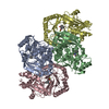
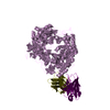
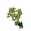
- PDB-6aca: Crystal structure of Mycobacterium tuberculosis Mfd at 3.6 A reso... -
+ Open data

- Basic information
| Entry | Database: PDB / ID: 6aca | ||||||
|---|---|---|---|---|---|---|---|
| Title | Crystal structure of Mycobacterium tuberculosis Mfd at 3.6 A resolution | ||||||
 Components Components | Mycobacterium tuberculosis Mfd | ||||||
 Keywords Keywords | HYDROLASE / Transcription repair coupling factor / Mfd / Transcription regulation / Transcription Coupled Nucleotide Excision Repair. | ||||||
| Function / homology |  Function and homology information Function and homology informationtranscription-coupled nucleotide-excision repair, DNA damage recognition / RNA polymerase core enzyme binding / DNA translocase activity / helicase activity / Hydrolases; Acting on acid anhydrides; Acting on acid anhydrides to facilitate cellular and subcellular movement / damaged DNA binding / hydrolase activity / regulation of DNA-templated transcription / DNA binding / ATP binding ...transcription-coupled nucleotide-excision repair, DNA damage recognition / RNA polymerase core enzyme binding / DNA translocase activity / helicase activity / Hydrolases; Acting on acid anhydrides; Acting on acid anhydrides to facilitate cellular and subcellular movement / damaged DNA binding / hydrolase activity / regulation of DNA-templated transcription / DNA binding / ATP binding / plasma membrane / cytosol Similarity search - Function | ||||||
| Biological species |  Mycobacterium tuberculosis H37Rv (bacteria) Mycobacterium tuberculosis H37Rv (bacteria) | ||||||
| Method |  X-RAY DIFFRACTION / SYNCHROTRON / MOLECULAR REPLACEMENT / Resolution: 3.6 Å X-RAY DIFFRACTION / SYNCHROTRON / MOLECULAR REPLACEMENT / Resolution: 3.6 Å | ||||||
 Authors Authors | Putta, S. / Fox, G.C. / Walsh, M.A. / Rao, D.N. / Nagaraja, V. / Natesh, R. | ||||||
| Funding support |  India, 1items India, 1items
| ||||||
 Citation Citation | Journal: To Be Published Title: Structural basis for nucleotide-mediated remodelling mechanism of Mycobacterium Mfd Authors: Putta, S. / Prabha, S. / Bhat, V. / Fox, G.C. / Walsh, M.A. / Rao, D.N. / Nagaraja, V. / Natesh, R. | ||||||
| History |
|
- Structure visualization
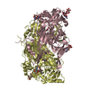
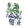
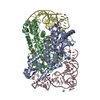
| Structure viewer | Molecule: MolmilJmol/JSmol |
|---|
- Downloads & links
-Download
| PDBx/mmCIF format | 6aca.cif.gz | 225.8 KB | Display | PDBx/mmCIF format |
|---|---|---|---|---|
| PDB format | pdb6aca.ent.gz | 172.7 KB | Display | PDB format |
| PDBx/mmJSON format | 6aca.json.gz | Tree view | PDBx/mmJSON format | |
| Others |  Other downloads Other downloads |
-Validation report
| Arichive directory | https://data.pdbj.org/pub/pdb/validation_reports/ac/6acaftp://data.pdbj.org/pub/pdb/validation_reports/ac/6aca | HTTPS FTP |
|---|
-Related structure data
| Related structure data |  9614C  6ac6C  6ac8SC  6acxC S: Starting model for refinement C: citing same article ( |
|---|---|
| Similar structure data |
-Links
 PDBj
PDBj- Assembly
| Deposited unit | 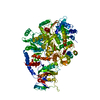
| ||||||||
|---|---|---|---|---|---|---|---|---|---|
| 1 |
| ||||||||
| Unit cell |
|
-Components
| #1: Protein | Mass: 135239.453 Da / Num. of mol.: 1 Source method: isolated from a genetically manipulated source Source: (gene. exp.) Mycobacterium tuberculosis H37Rv (bacteria)Strain: H37Rv / Gene: mfd / Plasmid: pETMtbMfd / Details (production host): MtbMfd gene cloned into pET28a / Production host: References: UniProt: P9WMQ5, Hydrolases; Acting on acid anhydrides; Acting on acid anhydrides to facilitate cellular and subcellular movement |
|---|---|
| #2: Water | ChemComp-HOH /  Mass: 18.015 Da / Num. of mol.: 16 / Source method: isolated from a natural source / Formula: H2O Mass: 18.015 Da / Num. of mol.: 16 / Source method: isolated from a natural source / Formula: H2O |
-Experimental details
-Experiment
| Experiment | Method: X-RAY DIFFRACTION / Number of used crystals: 1 |
|---|
- Sample preparation
| Crystal | Density Matthews: 3.47 Å3/Da / Density % sol: 64.6 % / Description: Cubic |
|---|---|
| Crystal grow | Temperature: 277 K / Method: vapor diffusion, hanging drop / pH: 7 Details: 100mM HEPES Sodium pH 7.0, 800mM Ammonium Formate, 20% PEG3350, 7.5mM MgCl2.6H2O PH range: 7.0-7.5 / Temp details: Rubarth Incubator |
-Data collection
| Diffraction | Mean temperature: 100 K / Ambient temp details: Cryo Stream |
|---|---|
| Diffraction source | Source: SYNCHROTRON / Site: Diamond  / Beamline: I03 / Wavelength: 0.9763 Å / Beamline: I03 / Wavelength: 0.9763 Å |
| Detector | Type: DECTRIS PILATUS3 6M / Detector: PIXEL / Date: Dec 12, 2015 |
| Radiation | Monochromator: Double Crystal / Protocol: SINGLE WAVELENGTH / Monochromatic (M) / Laue (L): M / Scattering type: x-ray |
| Radiation wavelength | Wavelength: 0.9763 Å / Relative weight: 1 |
| Reflection | Resolution: 3.6→39.64 Å / Num. obs: 22846 / % possible obs: 99.6 % / Redundancy: 8.7 % / Biso Wilson estimate: 110.5 Å2 / CC1/2: 0.998 / Rmerge(I) obs: 0.143 / Rpim(I) all: 0.052 / Rrim(I) all: 0.159 / Rsym value: 0.149 / Net I/σ(I): 8.7 |
| Reflection shell | Resolution: 3.6→3.89 Å / Redundancy: 8.7 % / Rmerge(I) obs: 1.006 / Mean I/σ(I) obs: 2.2 / Num. unique obs: 4588 / CC1/2: 0.381 / Rpim(I) all: 0.382 / Rrim(I) all: 1.141 / Rsym value: 1.069 / % possible all: 99.3 |
- Processing
| Software |
| |||||||||||||||||||||||||||||||||||||||||||||||||||||||||||||||
|---|---|---|---|---|---|---|---|---|---|---|---|---|---|---|---|---|---|---|---|---|---|---|---|---|---|---|---|---|---|---|---|---|---|---|---|---|---|---|---|---|---|---|---|---|---|---|---|---|---|---|---|---|---|---|---|---|---|---|---|---|---|---|---|---|
| Refinement | Method to determine structure: MOLECULAR REPLACEMENT Starting model: 6AC8 Resolution: 3.6→39.64 Å / Cross valid method: FREE R-VALUE / σ(F): 1.34 / Details: Phenix refinement
| |||||||||||||||||||||||||||||||||||||||||||||||||||||||||||||||
| Displacement parameters | Biso mean: 114.4 Å2 | |||||||||||||||||||||||||||||||||||||||||||||||||||||||||||||||
| Refinement step | Cycle: LAST / Resolution: 3.6→39.64 Å
| |||||||||||||||||||||||||||||||||||||||||||||||||||||||||||||||
| Refine LS restraints |
| |||||||||||||||||||||||||||||||||||||||||||||||||||||||||||||||
| LS refinement shell |
|
