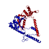
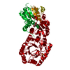
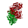
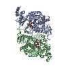
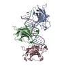
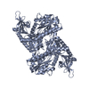
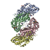
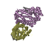
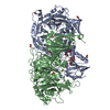
[English] 日本語
Yorodumi
- PDB-6a6g: Crystal structure of thermostable FiSufS-SufU complex from thermo... -

+
Open data


ID or keywords:

Loading...

-
Basic information

Entry
Database: PDB / ID: 6a6g
TitleCrystal structure of thermostable FiSufS-SufU complex from thermophilic Fervidobacterium Islandicum AW-1
Components
  • Cysteine desulfurase
  • Iron-sulfur cluster assembly scaffold protein NifU
KeywordsTRANSFERASE / Cysteine desulfurase (FiSufS) / Acceptor protein (FiSufE) / Thermophile / Suf gene cluster
Function / homology
Function and homology information


cysteine desulfurase / cysteine desulfurase activity / cysteine metabolic process / iron-sulfur cluster assembly / iron-sulfur cluster binding / pyridoxal phosphate binding / iron ion binding
Similarity search - Function
Sufe protein. Chain: A - #10 / Sufe protein. Chain: A / NIF system FeS cluster assembly, NifU, N-terminal / NifU-like N terminal domain / Cysteine desulfurase, SufS / Cysteine desulfurase / Aminotransferase class-V, pyridoxal-phosphate binding site / Aminotransferases class-V pyridoxal-phosphate attachment site. / Aminotransferase class V domain / Aminotransferase class-V ...Sufe protein. Chain: A - #10 / Sufe protein. Chain: A / NIF system FeS cluster assembly, NifU, N-terminal / NifU-like N terminal domain / Cysteine desulfurase, SufS / Cysteine desulfurase / Aminotransferase class-V, pyridoxal-phosphate binding site / Aminotransferases class-V pyridoxal-phosphate attachment site. / Aminotransferase class V domain / Aminotransferase class-V / Aspartate Aminotransferase; domain 2 / Type I PLP-dependent aspartate aminotransferase-like (Major domain) / Pyridoxal phosphate-dependent transferase, small domain / Pyridoxal phosphate-dependent transferase, major domain / Pyridoxal phosphate-dependent transferase / Alpha-Beta Complex / 3-Layer(aba) Sandwich / Alpha Beta
Similarity search - Domain/homology
DI(HYDROXYETHYL)ETHER / PYRIDOXAL-5'-PHOSPHATE / SUF system NifU family Fe-S cluster assembly protein / Cysteine desulfurase
Similarity search - Component
Biological speciesFervidobacterium islandicum (bacteria)
MethodX-RAY DIFFRACTION / SYNCHROTRON / MOLECULAR REPLACEMENT / Resolution: 2.49 Å
AuthorsDhanasingh, I. / Jin, H.S. / Lee, D.W. / Lee, S.H.
Funding support Korea, Republic Of, 2items
OrganizationGrant numberCountry
2014R1A2A2A01006765 Korea, Republic Of
2009-0064846 Korea, Republic Of
CitationJournal: Microb Biotechnol / Year: 2020
Title: The sulfur formation system mediating extracellular cysteine-cystine recycling in Fervidobacterium islandicum AW-1 is associated with keratin degradation.
Authors: Jin, H.S. / Dhanasingh, I. / Sung, J.Y. / La, J.W. / Lee, Y. / Lee, E.M. / Kang, Y. / Lee, D.Y. / Lee, S.H. / Lee, D.W.
History
DepositionJun 27, 2018Deposition site: PDBJ / Processing site: PDBJ
Revision 1.0Oct 9, 2019Provider: repository / Type: Initial release
Revision 1.1Nov 25, 2020Group: Database references / Derived calculations / Category: citation / pdbx_struct_conn_angle / struct_conn
Item: _citation.title / _pdbx_struct_conn_angle.ptnr1_auth_asym_id ..._citation.title / _pdbx_struct_conn_angle.ptnr1_auth_asym_id / _pdbx_struct_conn_angle.ptnr1_auth_comp_id / _pdbx_struct_conn_angle.ptnr1_auth_seq_id / _pdbx_struct_conn_angle.ptnr1_label_asym_id / _pdbx_struct_conn_angle.ptnr1_label_atom_id / _pdbx_struct_conn_angle.ptnr1_label_comp_id / _pdbx_struct_conn_angle.ptnr1_label_seq_id / _pdbx_struct_conn_angle.ptnr2_auth_asym_id / _pdbx_struct_conn_angle.ptnr2_auth_seq_id / _pdbx_struct_conn_angle.ptnr2_label_asym_id / _pdbx_struct_conn_angle.ptnr2_symmetry / _pdbx_struct_conn_angle.ptnr3_auth_asym_id / _pdbx_struct_conn_angle.ptnr3_auth_comp_id / _pdbx_struct_conn_angle.ptnr3_auth_seq_id / _pdbx_struct_conn_angle.ptnr3_label_asym_id / _pdbx_struct_conn_angle.ptnr3_label_atom_id / _pdbx_struct_conn_angle.ptnr3_label_comp_id / _pdbx_struct_conn_angle.ptnr3_label_seq_id / _pdbx_struct_conn_angle.value / _struct_conn.conn_type_id / _struct_conn.id / _struct_conn.pdbx_dist_value / _struct_conn.pdbx_leaving_atom_flag / _struct_conn.ptnr1_auth_asym_id / _struct_conn.ptnr1_auth_comp_id / _struct_conn.ptnr1_auth_seq_id / _struct_conn.ptnr1_label_asym_id / _struct_conn.ptnr1_label_atom_id / _struct_conn.ptnr1_label_comp_id / _struct_conn.ptnr1_label_seq_id / _struct_conn.ptnr2_auth_asym_id / _struct_conn.ptnr2_auth_comp_id / _struct_conn.ptnr2_auth_seq_id / _struct_conn.ptnr2_label_asym_id / _struct_conn.ptnr2_label_atom_id / _struct_conn.ptnr2_label_comp_id / _struct_conn.ptnr2_label_seq_id / _struct_conn.ptnr2_symmetry
Revision 1.2Dec 30, 2020Group: Database references / Category: citation / citation_author
Item: _citation.journal_abbrev / _citation.journal_id_CSD ..._citation.journal_abbrev / _citation.journal_id_CSD / _citation.journal_id_ISSN / _citation.pdbx_database_id_DOI / _citation.pdbx_database_id_PubMed / _citation.title / _citation.year
Revision 1.3Nov 22, 2023Group: Data collection / Database references ...Data collection / Database references / Derived calculations / Refinement description
Category: chem_comp_atom / chem_comp_bond ...chem_comp_atom / chem_comp_bond / database_2 / pdbx_initial_refinement_model / struct_conn
Item: _database_2.pdbx_DOI / _database_2.pdbx_database_accession / _struct_conn.pdbx_leaving_atom_flag

-
Structure visualization

Structure viewerMolecule:
MolmilJmol/JSmol

Downloads & links

-
Assembly

Deposited unit
A: Cysteine desulfurase
B: Cysteine desulfurase
C: Iron-sulfur cluster assembly scaffold protein NifU
D: Iron-sulfur cluster assembly scaffold protein NifU
hetero molecules


Theoretical massNumber of molelcules
Total (without water)128,05812
Polymers127,0634
Non-polymers9958
Water8,251458
1


  • Idetical with deposited unit
  • defined by author&software
  • Evidence: isothermal titration calorimetry
TypeNameSymmetry operationNumber
identity operation1_555x,y,z1
Buried area14030 Å2
ΔGint-167 kcal/mol
Surface area37510 Å2
MethodPISA
Unit cell
Length a, b, c (Å)75.265, 76.969, 192.520
Angle α, β, γ (deg.)90.00, 90.00, 90.00
Int Tables number19
Space group name H-MP212121

-
Components

-
Protein , 2 types, 4 molecules ABCD

#1: Protein Cysteine desulfurase / SufS


Mass: 47644.273 Da / Num. of mol.: 2
Source method: isolated from a genetically manipulated source
Source: (gene. exp.) Fervidobacterium islandicum (bacteria) / Gene: NA23_08315 / Production host: Escherichia coli BL21 (bacteria) / References: UniProt: A0A1B0VPZ3, cysteine desulfurase
#2: Protein Iron-sulfur cluster assembly scaffold protein NifU / SufE


Mass: 15887.457 Da / Num. of mol.: 2
Source method: isolated from a genetically manipulated source
Source: (gene. exp.) Fervidobacterium islandicum (bacteria) / Gene: NA23_08310 / Production host: Escherichia coli BL21 (bacteria) / References: UniProt: A0A1B0VLW5

-
Non-polymers , 5 types, 466 molecules

#3: Chemical ChemComp-PLP / PYRIDOXAL-5'-PHOSPHATE / VITAMIN B6 Phosphate


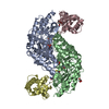
Mass: 247.142 Da / Num. of mol.: 2 / Source method: obtained synthetically / Formula: C8H10NO6P
#4: Chemical ChemComp-PEG / DI(HYDROXYETHYL)ETHER


Mass: 106.120 Da / Num. of mol.: 2 / Source method: obtained synthetically / Formula: C4H10O3
#5: Chemical ChemComp-GOL / GLYCEROL / GLYCERIN / PROPANE-1,2,3-TRIOL


Mass: 92.094 Da / Num. of mol.: 1 / Source method: obtained synthetically / Formula: C3H8O3
#6: Chemical ChemComp-ZN / ZINC ION


Mass: 65.409 Da / Num. of mol.: 3 / Source method: obtained synthetically / Formula: Zn
#7: Water ChemComp-HOH / water


Mass: 18.015 Da / Num. of mol.: 458 / Source method: isolated from a natural source / Formula: H2O

-
Experimental details

-
Experiment

ExperimentMethod: X-RAY DIFFRACTION / Number of used crystals: 1

-
Sample preparation

CrystalDensity Matthews: 2.19 Å3/Da / Density % sol: 43.95 %
Crystal growTemperature: 293 K / Method: vapor diffusion, hanging drop / Details: 0.2M lithium nitrate, PEG 3350, 0.1M HEPES, pH 7.0

-
Data collection

DiffractionMean temperature: 100 K
Diffraction sourceSource: SYNCHROTRON / Site: PAL/PLS / Beamline: 7A (6B, 6C1) / Wavelength: 0.979133 Å
DetectorType: ADSC QUANTUM 270 / Detector: CCD / Date: Oct 10, 2016
RadiationProtocol: SINGLE WAVELENGTH / Monochromatic (M) / Laue (L): M / Scattering type: x-ray
Radiation wavelengthWavelength: 0.979133 Å / Relative weight: 1
ReflectionResolution: 2.5→50 Å / Num. obs: 39603 / % possible obs: 98.95 % / Redundancy: 8.6 % / Rmerge(I) obs: 0.129 / Net I/σ(I): 7.46
Reflection shellResolution: 2.5→2.54 Å / Rmerge(I) obs: 0.435

-
Processing

Software
NameVersionClassification
REFMAC5.8.0218refinement
HKL-2000data reduction
HKL-2000data scaling
PHASERphasing
RefinementMethod to determine structure: MOLECULAR REPLACEMENT
Starting model: 6A6E and 6A6F
Resolution: 2.49→29.616 Å / Cor.coef. Fo:Fc: 0.965 / Cor.coef. Fo:Fc free: 0.927 / SU B: 8.114 / SU ML: 0.182 / Cross valid method: THROUGHOUT / ESU R: 2.011 / ESU R Free: 0.27 / Stereochemistry target values: MAXIMUM LIKELIHOOD / Details: HYDROGENS HAVE BEEN ADDED IN THE RIDING POSITIONS
RfactorNum. reflection% reflectionSelection details
Rfree0.2139 1912 4.8 %RANDOM
Rwork0.14633 ---
obs0.14962 37691 98.85 %-
Solvent computationIon probe radii: 0.8 Å / Shrinkage radii: 0.8 Å / VDW probe radii: 1.2 Å / Solvent model: MASK
Displacement parametersBiso mean: 38.12 Å2
Baniso -1Baniso -2Baniso -3
1--0 Å20 Å2-0 Å2
2--0 Å20 Å2
3---0 Å2
Refinement stepCycle: 1 / Resolution: 2.49→29.616 Å
ProteinNucleic acidLigandSolventTotal
Num. atoms8832 0 53 458 9343
Refine LS restraints
Refine-IDTypeDev idealDev ideal targetNumber
X-RAY DIFFRACTIONr_bond_refined_d0.0130.0199063
X-RAY DIFFRACTIONr_bond_other_d0.0010.028515
X-RAY DIFFRACTIONr_angle_refined_deg1.5111.96512224
X-RAY DIFFRACTIONr_angle_other_deg0.822319809
X-RAY DIFFRACTIONr_dihedral_angle_1_deg6.39551108
X-RAY DIFFRACTIONr_dihedral_angle_2_deg37.28425400
X-RAY DIFFRACTIONr_dihedral_angle_3_deg16.037151643
X-RAY DIFFRACTIONr_dihedral_angle_4_deg15.531526
X-RAY DIFFRACTIONr_chiral_restr0.0830.21376
X-RAY DIFFRACTIONr_gen_planes_refined0.0060.029894
X-RAY DIFFRACTIONr_gen_planes_other0.0010.021772
X-RAY DIFFRACTIONr_nbd_refined
X-RAY DIFFRACTIONr_nbd_other
X-RAY DIFFRACTIONr_nbtor_refined
X-RAY DIFFRACTIONr_nbtor_other
X-RAY DIFFRACTIONr_xyhbond_nbd_refined
X-RAY DIFFRACTIONr_xyhbond_nbd_other
X-RAY DIFFRACTIONr_metal_ion_refined
X-RAY DIFFRACTIONr_metal_ion_other
X-RAY DIFFRACTIONr_symmetry_vdw_refined
X-RAY DIFFRACTIONr_symmetry_vdw_other
X-RAY DIFFRACTIONr_symmetry_hbond_refined
X-RAY DIFFRACTIONr_symmetry_hbond_other
X-RAY DIFFRACTIONr_symmetry_metal_ion_refined
X-RAY DIFFRACTIONr_symmetry_metal_ion_other
X-RAY DIFFRACTIONr_mcbond_it2.4933.6154444
X-RAY DIFFRACTIONr_mcbond_other2.4883.6154443
X-RAY DIFFRACTIONr_mcangle_it3.8785.4135548
X-RAY DIFFRACTIONr_mcangle_other3.8795.4145549
X-RAY DIFFRACTIONr_scbond_it3.1514.0454619
X-RAY DIFFRACTIONr_scbond_other3.1514.0454619
X-RAY DIFFRACTIONr_scangle_it
X-RAY DIFFRACTIONr_scangle_other5.0515.8726677
X-RAY DIFFRACTIONr_long_range_B_refined6.96242.92410428
X-RAY DIFFRACTIONr_long_range_B_other6.96242.92810429
X-RAY DIFFRACTIONr_rigid_bond_restr
X-RAY DIFFRACTIONr_sphericity_free
X-RAY DIFFRACTIONr_sphericity_bonded
LS refinement shellResolution: 2.489→2.553 Å / Total num. of bins used: 20
RfactorNum. reflection% reflection
Rfree0.281 108 -
Rwork0.169 2464 -
obs--88.78 %

+
About Yorodumi

-
News

-
Feb 9, 2022. New format data for meta-information of EMDB entries

New format data for meta-information of EMDB entries

  • Version 3 of the EMDB header file is now the official format.
  • The previous official version 1.9 will be removed from the archive.

Related info.:EMDB header

External links:wwPDB to switch to version 3 of the EMDB data model

-
Aug 12, 2020. Covid-19 info

Covid-19 info

URL: https://pdbjlvh1.pdbj.org/emnavi/covid19.php

New page: Covid-19 featured information page in EM Navigator.

Related info.:Covid-19 info / Mar 5, 2020. Novel coronavirus structure data

+
Mar 5, 2020. Novel coronavirus structure data

Novel coronavirus structure data

Related info.:Yorodumi Speices / Aug 12, 2020. Covid-19 info

External links:COVID-19 featured content - PDBj / Molecule of the Month (242):Coronavirus Proteases

+
Jan 31, 2019. EMDB accession codes are about to change! (news from PDBe EMDB page)

EMDB accession codes are about to change! (news from PDBe EMDB page)

  • The allocation of 4 digits for EMDB accession codes will soon come to an end. Whilst these codes will remain in use, new EMDB accession codes will include an additional digit and will expand incrementally as the available range of codes is exhausted. The current 4-digit format prefixed with “EMD-” (i.e. EMD-XXXX) will advance to a 5-digit format (i.e. EMD-XXXXX), and so on. It is currently estimated that the 4-digit codes will be depleted around Spring 2019, at which point the 5-digit format will come into force.
  • The EM Navigator/Yorodumi systems omit the EMD- prefix.

Related info.:Q: What is EMD? / ID/Accession-code notation in Yorodumi/EM Navigator

External links:EMDB Accession Codes are Changing Soon! / Contact to PDBj

+
Jul 12, 2017. Major update of PDB

Major update of PDB

  • wwPDB released updated PDB data conforming to the new PDBx/mmCIF dictionary.
  • This is a major update changing the version number from 4 to 5, and with Remediation, in which all the entries are updated.
  • In this update, many items about electron microscopy experimental information are reorganized (e.g. em_software).
  • Now, EM Navigator and Yorodumi are based on the updated data.

External links:wwPDB Remediation / Enriched Model Files Conforming to OneDep Data Standards Now Available in the PDB FTP Archive

-
Yorodumi

Thousand views of thousand structures

  • Yorodumi is a browser for structure data from EMDB, PDB, SASBDB, etc.
  • This page is also the successor to EM Navigator detail page, and also detail information page/front-end page for Omokage search.
  • The word "yorodu" (or yorozu) is an old Japanese word meaning "ten thousand". "mi" (miru) is to see.

Related info.:EMDB / PDB / SASBDB / Comparison of 3 databanks / Yorodumi Search / Aug 31, 2016. New EM Navigator & Yorodumi / Yorodumi Papers / Jmol/JSmol / Function and homology information / Changes in new EM Navigator and Yorodumi

Read more