- PDB-6a58: Structure of histone demethylase REF6 -
+
Open data
ID or keywords:
Loading...
-
Basic information
Entry
Database: PDB / ID: 6a58
Title
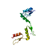
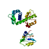
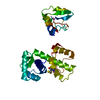
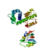
Structure of histone demethylase REF6
Components
Lysine-specific demethylase REF6
Keywords
DNA BINDING PROTEIN / Complex / histone demethylase REF6 / DNA / zinc finger
Function / homology
Function and homology information
regulation of ethylene-activated signaling pathway / abscisic acid catabolic process / release of seed from dormancy / positive regulation of lateral root development / response to diterpene / sugar mediated signaling pathway / protein localization to heterochromatin / heat acclimation / unidimensional cell growth / regulation of photoperiodism, flowering ...regulation of ethylene-activated signaling pathway / abscisic acid catabolic process / release of seed from dormancy / positive regulation of lateral root development / response to diterpene / sugar mediated signaling pathway / protein localization to heterochromatin / heat acclimation / unidimensional cell growth / regulation of photoperiodism, flowering / vegetative to reproductive phase transition of meristem / response to brassinosteroid / histone H3K27me2/H3K27me3 demethylase activity / systemic acquired resistance / ethylene-activated signaling pathway / leaf development / histone H3K4me/H3K4me2/H3K4me3 demethylase activity / response to abscisic acid / Oxidoreductases; Acting on paired donors, with incorporation or reduction of molecular oxygen; With 2-oxoglutarate as one donor, and incorporation of one atom of oxygen into each donor / Ino80 complex / abscisic acid-activated signaling pathway / response to mechanical stimulus / epigenetic regulation of gene expression / protein homooligomerization / response to heat / regulation of gene expression / sequence-specific DNA binding / chromatin remodeling / positive regulation of gene expression / regulation of DNA-templated transcription / chromatin / protein homodimerization activity / zinc ion binding / nucleus Similarity search - Function
JmjN domain / jmjN domain / JmjN domain profile. / Small domain found in the jumonji family of transcription factors / JmjC domain, hydroxylase / A domain family that is part of the cupin metalloenzyme superfamily. / JmjC domain / JmjC domain profile. / zinc finger / Zinc finger C2H2 type domain profile. ...JmjN domain / jmjN domain / JmjN domain profile. / Small domain found in the jumonji family of transcription factors / JmjC domain, hydroxylase / A domain family that is part of the cupin metalloenzyme superfamily. / JmjC domain / JmjC domain profile. / zinc finger / Zinc finger C2H2 type domain profile. / Zinc finger C2H2 superfamily / Zinc finger C2H2 type domain signature. / Zinc finger C2H2-type Similarity search - Domain/homology
Lysine-specificdemethylaseREF6 / Jumonji domain-containing protein 12 / Lysine-specific histone demethylase REF6 / Protein RELATIVE ...Jumonji domain-containing protein 12 / Lysine-specific histone demethylase REF6 / Protein RELATIVE OF EARLY FLOWERING 6
Mass: 16521.723 Da / Num. of mol.: 1 / Fragment: Ig gamma-1 chain C region Source method: isolated from a genetically manipulated source Source: (gene. exp.) Arabidopsis thaliana (thale cress) / Gene: REF6, JMJ12, PKDM9A, At3g48430, T29H11_50 / Production host: Escherichia coli (E. coli) References: UniProt: Q9STM3, Oxidoreductases; Acting on paired donors, with incorporation or reduction of molecular oxygen; With 2-oxoglutarate as one donor, and incorporation of one atom of oxygen into each donor
Resolution: 1.57→50 Å / Num. obs: 23425 / % possible obs: 99.7 % / Redundancy: 12.8 % / Rsym value: 0.057 / Net I/σ(I): 76.8
Reflection shell
Resolution: 1.57→1.6 Å / Num. unique obs: 1174 / Rsym value: 0.496 / % possible all: 99.1
-
Processing
Software
Name
Version
Classification
REFMAC
5.8.0189
refinement
HKL-2000
datareduction
HKL-2000
datascaling
HKL2Map
phasing
Refinement
Method to determine structure: MAD / Resolution: 1.57→50 Å / Cor.coef. Fo:Fc: 0.959 / Cor.coef. Fo:Fc free: 0.947 / SU B: 1.614 / SU ML: 0.057 / Cross valid method: THROUGHOUT / ESU R: 0.083 / ESU R Free: 0.082 / Details: HYDROGENS HAVE BEEN ADDED IN THE RIDING POSITIONS
Rfactor
Num. reflection
% reflection
Selection details
Rfree
0.23384
1187
5.1 %
RANDOM
Rwork
0.21085
-
-
-
obs
0.21199
22150
99.71 %
-
Solvent computation
Ion probe radii: 0.8 Å / Shrinkage radii: 0.8 Å / VDW probe radii: 1.2 Å
 Movie
Movie Controller


 Open data
 Basic information
 Components
 Keywords
 Function and homology information
 X-RAY DIFFRACTION /
  Authors
 China, 1items
  Citation
 Structure visualization
 Downloads & links
 Other downloads
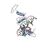


 PDBj
PDBj


 Assembly


 Mass: 65.409 Da / Num. of mol.: 4 / Source method: obtained synthetically / Formula: Zn
 Mass: 18.015 Da / Num. of mol.: 119 / Source method: isolated from a natural source / Formula: H2O
 Sample preparation
 Processing