 Movie
Movie Controller


+ Open data

- Basic information
| Entry | Database: PDB / ID: 6a2v | ||||||
|---|---|---|---|---|---|---|---|
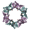
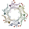
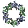
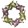
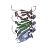
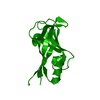
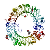
| Title | Crystal structure of Hcp protein | ||||||
 Components Components | Type VI secretion system tube protein Hcp | ||||||
 Keywords Keywords | STRUCTURAL PROTEIN / hemolysin coregulated protein / Type VI secretion system protein | ||||||
| Function / homology | Hcp1-like / Pnp Oxidase; Chain A / Roll / Mainly Beta / :  Function and homology information Function and homology information | ||||||
| Biological species |  Campylobacter jejuni subsp. jejuni (Campylobacter) Campylobacter jejuni subsp. jejuni (Campylobacter) | ||||||
| Method |  X-RAY DIFFRACTION / SYNCHROTRON / MOLECULAR REPLACEMENT / Resolution: 2.588 Å X-RAY DIFFRACTION / SYNCHROTRON / MOLECULAR REPLACEMENT / Resolution: 2.588 Å | ||||||
 Authors Authors | Jobichen, C. / Sivaraman, J. | ||||||
 Citation Citation | Journal: FEBS J. / Year: 2018 Title: Structural basis for the pathogenesis of Campylobacter jejuni Hcp1, a structural and effector protein of the Type VI Secretion System. Authors: Noreen, Z. / Jobichen, C. / Abbasi, R. / Seetharaman, J. / Sivaraman, J. / Bokhari, H. | ||||||
| History |
|
- Structure visualization
| Structure viewer | Molecule: MolmilJmol/JSmol |
|---|
- Downloads & links
-Download
| PDBx/mmCIF format | 6a2v.cif.gz | 189.7 KB | Display | PDBx/mmCIF format |
|---|---|---|---|---|
| PDB format | pdb6a2v.ent.gz | 150.8 KB | Display | PDB format |
| PDBx/mmJSON format | 6a2v.json.gz | Tree view | PDBx/mmJSON format | |
| Others |  Other downloads Other downloads |
-Validation report
| Arichive directory | https://data.pdbj.org/pub/pdb/validation_reports/a2/6a2vftp://data.pdbj.org/pub/pdb/validation_reports/a2/6a2v | HTTPS FTP |
|---|
-Related structure data
| Related structure data |  3he1S S: Starting model for refinement |
|---|---|
| Similar structure data |

-Links
 PDBj
PDBj- Assembly
| Deposited unit | 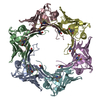
| ||||||||
|---|---|---|---|---|---|---|---|---|---|
| 1 |
| ||||||||
| Unit cell |
|
-Components
| #1: Protein | Mass: 18960.482 Da / Num. of mol.: 6 Source method: isolated from a genetically manipulated source Source: (gene. exp.) Campylobacter jejuni subsp. jejuni (Campylobacter)Gene: CV369_03455 / Production host: #2: Water | ChemComp-HOH / |  Mass: 18.015 Da / Num. of mol.: 98 / Source method: isolated from a natural source / Formula: H2O Mass: 18.015 Da / Num. of mol.: 98 / Source method: isolated from a natural source / Formula: H2OHas protein modification | Y | |
|---|
-Experimental details
-Experiment
| Experiment | Method: X-RAY DIFFRACTION / Number of used crystals: 1 |
|---|
- Sample preparation
| Crystal | Density Matthews: 3.09 Å3/Da / Density % sol: 64.07 % |
|---|---|
| Crystal grow | Temperature: 298 K / Method: vapor diffusion, hanging drop Details: Sodium acetate trihydrate, Tris HCl, Polyethylene glycol 3350 |
-Data collection
| Diffraction | Mean temperature: 100 K |
|---|---|
| Diffraction source | Source: SYNCHROTRON / Site: NSLS-II  / Beamline: 19-ID / Wavelength: 0.9789 Å / Beamline: 19-ID / Wavelength: 0.9789 Å |
| Detector | Type: DECTRIS EIGER X 16M / Detector: PIXEL / Date: Apr 26, 2018 |
| Radiation | Protocol: SINGLE WAVELENGTH / Monochromatic (M) / Laue (L): M / Scattering type: x-ray |
| Radiation wavelength | Wavelength: 0.9789 Å / Relative weight: 1 |
| Reflection | Resolution: 2.588→50 Å / Num. obs: 73986 / % possible obs: 87.5 % / Redundancy: 2.7 % / Rmerge(I) obs: 0.12 / Net I/σ(I): 15.4 |
| Reflection shell | Resolution: 2.588→2.64 Å / Rmerge(I) obs: 0.56 |
- Processing
| Software |
| ||||||||||||||||||||||||||||||||||||||||||||||||||||||||||||||||||||||||||||||||||||||||||||||||||||||||||||||||||||||||||||||||||||||||||||||||||||||||||||||||||||||||||||||||||||||||||||||||||||
|---|---|---|---|---|---|---|---|---|---|---|---|---|---|---|---|---|---|---|---|---|---|---|---|---|---|---|---|---|---|---|---|---|---|---|---|---|---|---|---|---|---|---|---|---|---|---|---|---|---|---|---|---|---|---|---|---|---|---|---|---|---|---|---|---|---|---|---|---|---|---|---|---|---|---|---|---|---|---|---|---|---|---|---|---|---|---|---|---|---|---|---|---|---|---|---|---|---|---|---|---|---|---|---|---|---|---|---|---|---|---|---|---|---|---|---|---|---|---|---|---|---|---|---|---|---|---|---|---|---|---|---|---|---|---|---|---|---|---|---|---|---|---|---|---|---|---|---|---|---|---|---|---|---|---|---|---|---|---|---|---|---|---|---|---|---|---|---|---|---|---|---|---|---|---|---|---|---|---|---|---|---|---|---|---|---|---|---|---|---|---|---|---|---|---|---|---|---|
| Refinement | Method to determine structure: MOLECULAR REPLACEMENT Starting model: 3HE1 Resolution: 2.588→19.968 Å / SU ML: 0.35 / Cross valid method: FREE R-VALUE / σ(F): 1.34 / Phase error: 28.3 Details: THE STRUCTURE FACTOR FILE CONTAINS FRIEDEL PAIRS IN I_PLUS/MINUS COLUMNS
| ||||||||||||||||||||||||||||||||||||||||||||||||||||||||||||||||||||||||||||||||||||||||||||||||||||||||||||||||||||||||||||||||||||||||||||||||||||||||||||||||||||||||||||||||||||||||||||||||||||
| Solvent computation | Shrinkage radii: 0.9 Å / VDW probe radii: 1.11 Å | ||||||||||||||||||||||||||||||||||||||||||||||||||||||||||||||||||||||||||||||||||||||||||||||||||||||||||||||||||||||||||||||||||||||||||||||||||||||||||||||||||||||||||||||||||||||||||||||||||||
| Refinement step | Cycle: LAST / Resolution: 2.588→19.968 Å
| ||||||||||||||||||||||||||||||||||||||||||||||||||||||||||||||||||||||||||||||||||||||||||||||||||||||||||||||||||||||||||||||||||||||||||||||||||||||||||||||||||||||||||||||||||||||||||||||||||||
| Refine LS restraints |
| ||||||||||||||||||||||||||||||||||||||||||||||||||||||||||||||||||||||||||||||||||||||||||||||||||||||||||||||||||||||||||||||||||||||||||||||||||||||||||||||||||||||||||||||||||||||||||||||||||||
| LS refinement shell |
|
