 Movie
Movie Controller

[English] 日本語
 Yorodumi
- PDB-6a0r: Homoserine dehydrogenase from Thermus thermophilus HB8 unliganded form -
+ Open data

- Basic information
| Entry | Database: PDB / ID: 6a0r | ||||||
|---|---|---|---|---|---|---|---|
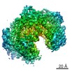
| Title | Homoserine dehydrogenase from Thermus thermophilus HB8 unliganded form | ||||||
 Components Components | Homoserine dehydrogenase | ||||||
 Keywords Keywords | OXIDOREDUCTASE / nad-dependent / dehydrogenase | ||||||
| Function / homology |  Function and homology information Function and homology informationhomoserine dehydrogenase / homoserine dehydrogenase activity / L-threonine biosynthetic process / : / NADP binding / metal ion binding Similarity search - Function | ||||||
| Biological species |   Thermus thermophilus HB8 (bacteria) Thermus thermophilus HB8 (bacteria) | ||||||
| Method |  X-RAY DIFFRACTION / SYNCHROTRON / MOLECULAR REPLACEMENT / Resolution: 1.83 Å X-RAY DIFFRACTION / SYNCHROTRON / MOLECULAR REPLACEMENT / Resolution: 1.83 Å | ||||||
 Authors Authors | Akai, S. / Ikushiro, H. / Sawai, T. / Yano, T. / Kamiya, N. / Miyahara, I. | ||||||
 Citation Citation | Journal: J. Biochem. / Year: 2019 Title: The crystal structure of homoserine dehydrogenase complexed with l-homoserine and NADPH in a closed form Authors: Akai, S. / Ikushiro, H. / Sawai, T. / Yano, T. / Kamiya, N. / Miyahara, I. | ||||||
| History |
|
- Structure visualization
| Structure viewer | Molecule: MolmilJmol/JSmol |
|---|
- Downloads & links
-Download
| PDBx/mmCIF format | 6a0r.cif.gz | 167.3 KB | Display | PDBx/mmCIF format |
|---|---|---|---|---|
| PDB format | pdb6a0r.ent.gz | 130.7 KB | Display | PDB format |
| PDBx/mmJSON format | 6a0r.json.gz | Tree view | PDBx/mmJSON format | |
| Others |  Other downloads Other downloads |
-Validation report
| Arichive directory | https://data.pdbj.org/pub/pdb/validation_reports/a0/6a0rftp://data.pdbj.org/pub/pdb/validation_reports/a0/6a0r | HTTPS FTP |
|---|
-Related structure data
| Related structure data |  6a0sC 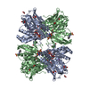 6a0tC 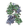 6a0uC 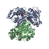 5xdfS S: Starting model for refinement C: citing same article ( |
|---|---|
| Similar structure data |


-Links
 PDBj
PDBj

- Assembly
| Deposited unit | 
| ||||||||
|---|---|---|---|---|---|---|---|---|---|
| 1 | 
| ||||||||
| Unit cell |
|
-Components
-Protein , 1 types, 2 molecules BA
| #1: Protein | Mass: 35529.820 Da / Num. of mol.: 2 Source method: isolated from a genetically manipulated source Source: (gene. exp.) Thermus thermophilus HB8 (bacteria) / Strain: HB8 / Gene: TTHA0489 / Production host: |
|---|
-Non-polymers , 6 types, 783 molecules 


| #2: Chemical |  Mass: 22.990 Da / Num. of mol.: 2 / Source method: obtained synthetically / Formula: Na Mass: 22.990 Da / Num. of mol.: 2 / Source method: obtained synthetically / Formula: Na#3: Chemical | ChemComp-FMT /  Mass: 46.025 Da / Num. of mol.: 17 / Source method: obtained synthetically / Formula: CH2O2 Mass: 46.025 Da / Num. of mol.: 17 / Source method: obtained synthetically / Formula: CH2O2#4: Chemical |  Mass: 221.317 Da / Num. of mol.: 2 / Source method: obtained synthetically / Formula: C9H19NO3S / Comment: pH buffer*YM Mass: 221.317 Da / Num. of mol.: 2 / Source method: obtained synthetically / Formula: C9H19NO3S / Comment: pH buffer*YM#5: Chemical |  Mass: 92.094 Da / Num. of mol.: 2 / Source method: obtained synthetically / Formula: C3H8O3 Mass: 92.094 Da / Num. of mol.: 2 / Source method: obtained synthetically / Formula: C3H8O3#6: Chemical | ChemComp-UNL / | Mass: 178.228 Da / Num. of mol.: 1 / Source method: obtained synthetically #7: Water | ChemComp-HOH / | Mass: 18.015 Da / Num. of mol.: 759 / Source method: isolated from a natural source / Formula: H2O |
|---|
-Details
| Nonpolymer details | Authors state that they do not use 4-~{tert}-butylbenzoic acid or the derivatives as a ...Authors state that they do not use 4-~{tert}-butylbenzoic acid or the derivatives as a crystallization solution. Therefore, even though the electron density map is clearly 4-~{tert}-butylbenzoic acid, the ligand is assigned to 'unknown ligand (UNL)' |
|---|
-Experimental details
-Experiment
| Experiment | Method: X-RAY DIFFRACTION / Number of used crystals: 1 |
|---|
- Sample preparation
| Crystal | Density Matthews: 4.17 Å3/Da / Density % sol: 70.51 % |
|---|---|
| Crystal grow | Temperature: 298 K / Method: vapor diffusion, hanging drop / Details: Sodium formate, CAPS pH 10.0 |
-Data collection
| Diffraction | Mean temperature: 100 K |
|---|---|
| Diffraction source | Source: SYNCHROTRON / Site: Photon Factory  / Beamline: AR-NW12A / Wavelength: 1 Å / Beamline: AR-NW12A / Wavelength: 1 Å |
| Detector | Type: ADSC QUANTUM 210 / Detector: CCD / Date: Jun 22, 2014 |
| Radiation | Protocol: SINGLE WAVELENGTH / Monochromatic (M) / Laue (L): M / Scattering type: x-ray |
| Radiation wavelength | Wavelength: 1 Å / Relative weight: 1 |
| Reflection | Resolution: 1.83→50 Å / Num. obs: 104929 / % possible obs: 100 % / Redundancy: 8.4 % / Rmerge(I) obs: 0.114 / Net I/σ(I): 48.7 |
| Reflection shell | Resolution: 1.83→1.86 Å / Redundancy: 8.4 % / Rmerge(I) obs: 0.359 / Mean I/σ(I) obs: 7 / % possible all: 100 |
- Processing
| Software |
| ||||||||||||||||||||||||||||||||||||||||||||||||||||||||||||||||||||||||||||||||||||||||||||||||||||||||||||||||||||||||||||||||||||||||||||||||||||||||||||||||||||||||||||||||||||||
|---|---|---|---|---|---|---|---|---|---|---|---|---|---|---|---|---|---|---|---|---|---|---|---|---|---|---|---|---|---|---|---|---|---|---|---|---|---|---|---|---|---|---|---|---|---|---|---|---|---|---|---|---|---|---|---|---|---|---|---|---|---|---|---|---|---|---|---|---|---|---|---|---|---|---|---|---|---|---|---|---|---|---|---|---|---|---|---|---|---|---|---|---|---|---|---|---|---|---|---|---|---|---|---|---|---|---|---|---|---|---|---|---|---|---|---|---|---|---|---|---|---|---|---|---|---|---|---|---|---|---|---|---|---|---|---|---|---|---|---|---|---|---|---|---|---|---|---|---|---|---|---|---|---|---|---|---|---|---|---|---|---|---|---|---|---|---|---|---|---|---|---|---|---|---|---|---|---|---|---|---|---|---|---|
| Refinement | Method to determine structure: MOLECULAR REPLACEMENT Starting model: 5XDF Resolution: 1.83→20 Å / Cor.coef. Fo:Fc: 0.974 / Cor.coef. Fo:Fc free: 0.964 / Cross valid method: THROUGHOUT / ESU R: 0.079 / ESU R Free: 0.082 / Stereochemistry target values: MAXIMUM LIKELIHOOD / Details: HYDROGENS HAVE BEEN ADDED IN THE RIDING POSITIONS
| ||||||||||||||||||||||||||||||||||||||||||||||||||||||||||||||||||||||||||||||||||||||||||||||||||||||||||||||||||||||||||||||||||||||||||||||||||||||||||||||||||||||||||||||||||||||
| Solvent computation | Ion probe radii: 0.8 Å / Shrinkage radii: 0.8 Å / VDW probe radii: 1.2 Å / Solvent model: MASK | ||||||||||||||||||||||||||||||||||||||||||||||||||||||||||||||||||||||||||||||||||||||||||||||||||||||||||||||||||||||||||||||||||||||||||||||||||||||||||||||||||||||||||||||||||||||
| Displacement parameters | Biso mean: 28.115 Å2
| ||||||||||||||||||||||||||||||||||||||||||||||||||||||||||||||||||||||||||||||||||||||||||||||||||||||||||||||||||||||||||||||||||||||||||||||||||||||||||||||||||||||||||||||||||||||
| Refinement step | Cycle: 1 / Resolution: 1.83→20 Å
| ||||||||||||||||||||||||||||||||||||||||||||||||||||||||||||||||||||||||||||||||||||||||||||||||||||||||||||||||||||||||||||||||||||||||||||||||||||||||||||||||||||||||||||||||||||||
| Refine LS restraints |
|