 Movie
Movie Controller


+ Open data

- Basic information
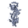
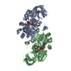
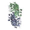
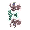
| Entry | Database: PDB / ID: 6a0q | |||||||||
|---|---|---|---|---|---|---|---|---|---|---|
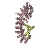
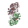
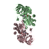
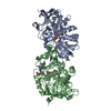
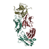
| Title | The crystal structure of Lpg2622_E64 complex | |||||||||
 Components Components | Lpg2622 | |||||||||
 Keywords Keywords | HYDROLASE / Cysteine protease | |||||||||
| Function / homology | Papain-like cysteine peptidase superfamily / Chem-E64 / Cysteine protease Function and homology information Function and homology information | |||||||||
| Biological species |  Legionella pneumophila subsp. pneumophila str. Philadelphia 1 (bacteria) Legionella pneumophila subsp. pneumophila str. Philadelphia 1 (bacteria) | |||||||||
| Method |  X-RAY DIFFRACTION / SYNCHROTRON / MOLECULAR REPLACEMENT / Resolution: 2.2 Å X-RAY DIFFRACTION / SYNCHROTRON / MOLECULAR REPLACEMENT / Resolution: 2.2 Å | |||||||||
 Authors Authors | Gong, X. / Ge, H. | |||||||||
| Funding support |  China, 2items China, 2items
| |||||||||
 Citation Citation | Journal: FEBS Lett. / Year: 2018 Title: Structural characterization of the hypothetical protein Lpg2622, a new member of the C1 family peptidases from Legionella pneumophila Authors: Gong, X. / Zhao, X. / Zhang, W. / Wang, J. / Chen, X. / Hameed, M.F. / Zhang, N. / Ge, H. | |||||||||
| History |
|
- Structure visualization
| Structure viewer | Molecule: MolmilJmol/JSmol |
|---|
- Downloads & links
-Download
| PDBx/mmCIF format | 6a0q.cif.gz | 137.6 KB | Display | PDBx/mmCIF format |
|---|---|---|---|---|
| PDB format | pdb6a0q.ent.gz | 108.5 KB | Display | PDB format |
| PDBx/mmJSON format | 6a0q.json.gz | Tree view | PDBx/mmJSON format | |
| Others |  Other downloads Other downloads |
-Validation report
| Summary document | 6a0q_validation.pdf.gz | 707.3 KB | Display | wwPDB validaton report |
|---|---|---|---|---|
| Full document | 6a0q_full_validation.pdf.gz | 727.6 KB | Display | |
| Data in XML | 6a0q_validation.xml.gz | 27.9 KB | Display | |
| Data in CIF | 6a0q_validation.cif.gz | 38.2 KB | Display | |
| Arichive directory | https://data.pdbj.org/pub/pdb/validation_reports/a0/6a0qftp://data.pdbj.org/pub/pdb/validation_reports/a0/6a0q | HTTPS FTP |
-Related structure data


-Links
 PDBj
PDBj- Assembly
| Deposited unit | 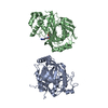
| ||||||||
|---|---|---|---|---|---|---|---|---|---|
| 1 | 
| ||||||||
| 2 | 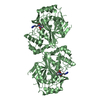
| ||||||||
| Unit cell |
| ||||||||
| Components on special symmetry positions |
|
-Components
| #1: Protein | Mass: 38081.855 Da / Num. of mol.: 2 / Fragment: UNP residues 20-353 Source method: isolated from a genetically manipulated source Source: (gene. exp.) Legionella pneumophila subsp. pneumophila str. Philadelphia 1 (bacteria)Strain: Philadelphia 1 / Gene: lpg2622 / Production host: #2: Chemical | ChemComp-E64 / |   Mass: 360.429 Da / Num. of mol.: 1 / Source method: obtained synthetically / Formula: C15H30N5O5 Mass: 360.429 Da / Num. of mol.: 1 / Source method: obtained synthetically / Formula: C15H30N5O5#3: Water | ChemComp-HOH / |  Mass: 18.015 Da / Num. of mol.: 123 / Source method: isolated from a natural source / Formula: H2O Mass: 18.015 Da / Num. of mol.: 123 / Source method: isolated from a natural source / Formula: H2O |
|---|
-Experimental details
-Experiment
| Experiment | Method: X-RAY DIFFRACTION / Number of used crystals: 1 |
|---|
- Sample preparation
| Crystal | Density Matthews: 3.47 Å3/Da / Density % sol: 64.53 % |
|---|---|
| Crystal grow | Temperature: 287 K / Method: vapor diffusion, hanging drop / Details: 0.4 M Sodium acetate trihydrate, pH 4.6 |
-Data collection
| Diffraction | Mean temperature: 100 K |
|---|---|
| Diffraction source | Source: SYNCHROTRON / Site: SSRF / Beamline: BL17U1 / Wavelength: 0.97916 Å |
| Detector | Type: ADSC QUANTUM 315r / Detector: CCD / Date: Dec 23, 2016 |
| Radiation | Protocol: SINGLE WAVELENGTH / Monochromatic (M) / Laue (L): M / Scattering type: x-ray |
| Radiation wavelength | Wavelength: 0.97916 Å / Relative weight: 1 |
| Reflection | Resolution: 2.2→50 Å / Num. obs: 54772 / % possible obs: 100 % / Redundancy: 21.7 % / Net I/σ(I): 27.2 |
| Reflection shell | Resolution: 2.2→2.24 Å |
- Processing
| Software |
| ||||||||||||||||||||||||||||||||||||||||||||||||||||||||||||||||||||||||||||||||||||||||||||||||||||||||||||||||||||||||||||||||||||||||||||||||||||||||||||||||||||||||||||||||||||||
|---|---|---|---|---|---|---|---|---|---|---|---|---|---|---|---|---|---|---|---|---|---|---|---|---|---|---|---|---|---|---|---|---|---|---|---|---|---|---|---|---|---|---|---|---|---|---|---|---|---|---|---|---|---|---|---|---|---|---|---|---|---|---|---|---|---|---|---|---|---|---|---|---|---|---|---|---|---|---|---|---|---|---|---|---|---|---|---|---|---|---|---|---|---|---|---|---|---|---|---|---|---|---|---|---|---|---|---|---|---|---|---|---|---|---|---|---|---|---|---|---|---|---|---|---|---|---|---|---|---|---|---|---|---|---|---|---|---|---|---|---|---|---|---|---|---|---|---|---|---|---|---|---|---|---|---|---|---|---|---|---|---|---|---|---|---|---|---|---|---|---|---|---|---|---|---|---|---|---|---|---|---|---|---|
| Refinement | Method to determine structure: MOLECULAR REPLACEMENT / Resolution: 2.2→50 Å / Cor.coef. Fo:Fc: 0.95 / Cor.coef. Fo:Fc free: 0.93 / Cross valid method: THROUGHOUT / ESU R: 0.186 / ESU R Free: 0.17 / Stereochemistry target values: MAXIMUM LIKELIHOOD / Details: HYDROGENS HAVE BEEN ADDED IN THE RIDING POSITIONS
| ||||||||||||||||||||||||||||||||||||||||||||||||||||||||||||||||||||||||||||||||||||||||||||||||||||||||||||||||||||||||||||||||||||||||||||||||||||||||||||||||||||||||||||||||||||||
| Solvent computation | Ion probe radii: 0.8 Å / Shrinkage radii: 0.8 Å / VDW probe radii: 1.2 Å / Solvent model: MASK | ||||||||||||||||||||||||||||||||||||||||||||||||||||||||||||||||||||||||||||||||||||||||||||||||||||||||||||||||||||||||||||||||||||||||||||||||||||||||||||||||||||||||||||||||||||||
| Displacement parameters | Biso mean: 43.083 Å2
| ||||||||||||||||||||||||||||||||||||||||||||||||||||||||||||||||||||||||||||||||||||||||||||||||||||||||||||||||||||||||||||||||||||||||||||||||||||||||||||||||||||||||||||||||||||||
| Refinement step | Cycle: 1 / Resolution: 2.2→50 Å
| ||||||||||||||||||||||||||||||||||||||||||||||||||||||||||||||||||||||||||||||||||||||||||||||||||||||||||||||||||||||||||||||||||||||||||||||||||||||||||||||||||||||||||||||||||||||
| Refine LS restraints |
|