 Movie
Movie Controller

[English] 日本語
 Yorodumi
- PDB-5zqs: Crystal structure of beta-xylosidase mutant (E186Q/F503Y) from Ba... -
+ Open data

- Basic information
| Entry | Database: PDB / ID: 5zqs | |||||||||
|---|---|---|---|---|---|---|---|---|---|---|
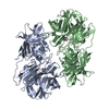
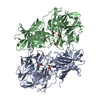
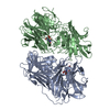
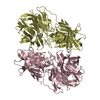
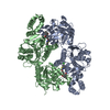
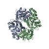
| Title | Crystal structure of beta-xylosidase mutant (E186Q/F503Y) from Bacillus pumilus | |||||||||
 Components Components | Beta-xylosidase | |||||||||
 Keywords Keywords | HYDROLASE / xylobiose hydrolysis | |||||||||
| Function / homology |  Function and homology information Function and homology informationxylan 1,4-beta-xylosidase / xylan 1,4-beta-xylosidase activity / xylan catabolic process Similarity search - Function | |||||||||
| Biological species |  | |||||||||
| Method |  X-RAY DIFFRACTION / SYNCHROTRON / MOLECULAR REPLACEMENT / Resolution: 1.782 Å X-RAY DIFFRACTION / SYNCHROTRON / MOLECULAR REPLACEMENT / Resolution: 1.782 Å | |||||||||
 Authors Authors | Ha, N.C. / Hong, S. / Jo, I. | |||||||||
 Citation Citation | Journal: Biochem. Biophys. Res. Commun. / Year: 2018 Title: Structure-based protein engineering of bacterial beta-xylosidase to increase the production yield of xylobiose from xylose Authors: Hong, S. / Kyung, M. / Jo, I. / Kim, Y.R. / Ha, N.C. | |||||||||
| History |
|
- Structure visualization
| Structure viewer | Molecule: MolmilJmol/JSmol |
|---|
- Downloads & links
-Download
| PDBx/mmCIF format | 5zqs.cif.gz | 237.4 KB | Display | PDBx/mmCIF format |
|---|---|---|---|---|
| PDB format | pdb5zqs.ent.gz | 188.3 KB | Display | PDB format |
| PDBx/mmJSON format | 5zqs.json.gz | Tree view | PDBx/mmJSON format | |
| Others |  Other downloads Other downloads |
-Validation report
| Arichive directory | https://data.pdbj.org/pub/pdb/validation_reports/zq/5zqsftp://data.pdbj.org/pub/pdb/validation_reports/zq/5zqs | HTTPS FTP |
|---|
-Related structure data
| Related structure data |  5zqjSC  5zqxC S: Starting model for refinement C: citing same article ( |
|---|---|
| Similar structure data |


-Links
 PDBj
PDBj- Assembly
| Deposited unit | 
| ||||||||
|---|---|---|---|---|---|---|---|---|---|
| 1 |
| ||||||||
| Unit cell |
|
-Components
| #1: Protein | Mass: 62105.488 Da / Num. of mol.: 2 / Mutation: E186Q, F503Y Source method: isolated from a genetically manipulated source Details: Electron map was not observed between 272-274. / Source: (gene. exp.) #2: Polysaccharide | 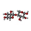  Source method: isolated from a genetically manipulated source Details: oligosaccharide / References: 4beta-beta-xylobiose #3: Sugar | ChemComp-XYP / |   Type: D-saccharide, beta linking / Mass: 150.130 Da / Num. of mol.: 1 Type: D-saccharide, beta linking / Mass: 150.130 Da / Num. of mol.: 1Source method: isolated from a genetically manipulated source Formula: C5H10O5 #4: Water | ChemComp-HOH / |  Mass: 18.015 Da / Num. of mol.: 555 / Source method: isolated from a natural source / Formula: H2O Mass: 18.015 Da / Num. of mol.: 555 / Source method: isolated from a natural source / Formula: H2O |
|---|
-Experimental details
-Experiment
| Experiment | Method: X-RAY DIFFRACTION / Number of used crystals: 1 |
|---|
- Sample preparation
| Crystal | Density Matthews: 2.14 Å3/Da / Density % sol: 42.51 % |
|---|---|
| Crystal grow | Temperature: 287 K / Method: vapor diffusion, hanging drop / pH: 7.5 Details: 0.05 M HEPES (pH 7.5), 20% (w/v) PEG 3350, 1% (w/v) tryptone, and 30 mM xylobiose |
-Data collection
| Diffraction | Mean temperature: 100 K |
|---|---|
| Diffraction source | Source: SYNCHROTRON / Site: PAL/PLS  / Beamline: 5C (4A) / Wavelength: 0.9794 Å / Beamline: 5C (4A) / Wavelength: 0.9794 Å |
| Detector | Type: ADSC QUANTUM 315 / Detector: CCD / Date: Dec 12, 2017 |
| Radiation | Protocol: SINGLE WAVELENGTH / Monochromatic (M) / Laue (L): M / Scattering type: x-ray |
| Radiation wavelength | Wavelength: 0.9794 Å / Relative weight: 1 |
| Reflection | Resolution: 1.78→50 Å / Num. obs: 95015 / % possible obs: 96.1 % / Observed criterion σ(I): 0 / Redundancy: 4.7 % / Rmerge(I) obs: 0.085 / Rpim(I) all: 0.038 / Rrim(I) all: 0.094 / Χ2: 0.87 / Net I/σ(I): 13.86 |
| Reflection shell | Resolution: 1.78→1.81 Å / Redundancy: 2.6 % / Rmerge(I) obs: 0.466 / Mean I/σ(I) obs: 1.97 / Num. unique obs: 4377 / CC1/2: 0.268 / Rpim(I) all: 0.298 / Rrim(I) all: 0.558 / Χ2: 0.763 / % possible all: 88.8 |
- Processing
| Software |
| |||||||||||||||||||||||||||||||||||||||||||||||||||||||||||||||||||||||||||||||||||||||||||||||||||||||||||||||||||||||||||||||||||||||||||||||||||||||||||||||||||||||||||||||||||||||||||||||||||||||||||||||||||||||||
|---|---|---|---|---|---|---|---|---|---|---|---|---|---|---|---|---|---|---|---|---|---|---|---|---|---|---|---|---|---|---|---|---|---|---|---|---|---|---|---|---|---|---|---|---|---|---|---|---|---|---|---|---|---|---|---|---|---|---|---|---|---|---|---|---|---|---|---|---|---|---|---|---|---|---|---|---|---|---|---|---|---|---|---|---|---|---|---|---|---|---|---|---|---|---|---|---|---|---|---|---|---|---|---|---|---|---|---|---|---|---|---|---|---|---|---|---|---|---|---|---|---|---|---|---|---|---|---|---|---|---|---|---|---|---|---|---|---|---|---|---|---|---|---|---|---|---|---|---|---|---|---|---|---|---|---|---|---|---|---|---|---|---|---|---|---|---|---|---|---|---|---|---|---|---|---|---|---|---|---|---|---|---|---|---|---|---|---|---|---|---|---|---|---|---|---|---|---|---|---|---|---|---|---|---|---|---|---|---|---|---|---|---|---|---|---|---|---|---|
| Refinement | Method to determine structure: MOLECULAR REPLACEMENT Starting model: 5ZQJ Resolution: 1.782→35.633 Å / SU ML: 0.17 / Cross valid method: FREE R-VALUE / σ(F): 1.53 / Phase error: 20.33
| |||||||||||||||||||||||||||||||||||||||||||||||||||||||||||||||||||||||||||||||||||||||||||||||||||||||||||||||||||||||||||||||||||||||||||||||||||||||||||||||||||||||||||||||||||||||||||||||||||||||||||||||||||||||||
| Solvent computation | Shrinkage radii: 0.9 Å / VDW probe radii: 1.11 Å | |||||||||||||||||||||||||||||||||||||||||||||||||||||||||||||||||||||||||||||||||||||||||||||||||||||||||||||||||||||||||||||||||||||||||||||||||||||||||||||||||||||||||||||||||||||||||||||||||||||||||||||||||||||||||
| Refinement step | Cycle: LAST / Resolution: 1.782→35.633 Å
| |||||||||||||||||||||||||||||||||||||||||||||||||||||||||||||||||||||||||||||||||||||||||||||||||||||||||||||||||||||||||||||||||||||||||||||||||||||||||||||||||||||||||||||||||||||||||||||||||||||||||||||||||||||||||
| Refine LS restraints |
| |||||||||||||||||||||||||||||||||||||||||||||||||||||||||||||||||||||||||||||||||||||||||||||||||||||||||||||||||||||||||||||||||||||||||||||||||||||||||||||||||||||||||||||||||||||||||||||||||||||||||||||||||||||||||
| LS refinement shell |
|
