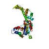
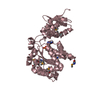
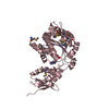
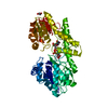
[English] 日本語
Yorodumi
- PDB-5zqm: Crystal structure of human katanin AAA ATPase domain complex with... -

+
Open data


ID or keywords:

Loading...

-
Basic information

Entry
Database: PDB / ID: 5zqm
TitleCrystal structure of human katanin AAA ATPase domain complex with ATPgammaS
ComponentsKatanin p60 ATPase-containing subunit A1
KeywordsHYDROLASE / Katanin p60 / AAA ATPase
Function / homology
Function and homology information


katanin complex / microtubule-severing ATPase / microtubule severing ATPase activity / microtubule severing / mitotic spindle pole / cytoplasmic microtubule organization / spindle / spindle pole / microtubule cytoskeleton / midbody ...katanin complex / microtubule-severing ATPase / microtubule severing ATPase activity / microtubule severing / mitotic spindle pole / cytoplasmic microtubule organization / spindle / spindle pole / microtubule cytoskeleton / midbody / microtubule binding / microtubule / protein heterodimerization activity / cell division / centrosome / ATP hydrolysis activity / ATP binding / cytoplasm
Similarity search - Function
: / : / Katanin p60 subunit A1, MIT domain / Katanin p60 subunit A1 / Vps4 oligomerisation, C-terminal / : / Vps4 C terminal oligomerisation domain / AAA ATPase, AAA+ lid domain / AAA+ lid domain / ATPase, AAA-type, conserved site ...: / : / Katanin p60 subunit A1, MIT domain / Katanin p60 subunit A1 / Vps4 oligomerisation, C-terminal / : / Vps4 C terminal oligomerisation domain / AAA ATPase, AAA+ lid domain / AAA+ lid domain / ATPase, AAA-type, conserved site / AAA-protein family signature. / ATPase family associated with various cellular activities (AAA) / ATPase, AAA-type, core / P-loop containing nucleotide triphosphate hydrolases / ATPases associated with a variety of cellular activities / AAA+ ATPase domain / Rossmann fold / P-loop containing nucleoside triphosphate hydrolase / 3-Layer(aba) Sandwich / Alpha Beta
Similarity search - Domain/homology
PHOSPHOTHIOPHOSPHORIC ACID-ADENYLATE ESTER / Katanin p60 ATPase-containing subunit A1
Similarity search - Component
Biological speciesHomo sapiens (human)
MethodX-RAY DIFFRACTION / SYNCHROTRON / MOLECULAR REPLACEMENT / Resolution: 2.9 Å
AuthorsKim, E.E. / Shin, S.C.
Funding support Korea, Republic Of, 1items
OrganizationGrant numberCountry
Korea, Republic Of
CitationJournal: Cell Rep / Year: 2019
Title: Structural and Molecular Basis for Katanin-Mediated Severing of Glutamylated Microtubules.
Authors: Shin, S.C. / Im, S.K. / Jang, E.H. / Jin, K.S. / Hur, E.M. / Kim, E.E.
History
DepositionApr 19, 2018Deposition site: PDBJ / Processing site: PDBJ
Revision 1.0Feb 27, 2019Provider: repository / Type: Initial release
Revision 1.1Nov 22, 2023Group: Data collection / Database references / Refinement description
Category: chem_comp_atom / chem_comp_bond ...chem_comp_atom / chem_comp_bond / database_2 / pdbx_initial_refinement_model
Item: _database_2.pdbx_DOI / _database_2.pdbx_database_accession

-
Structure visualization

Structure viewerMolecule:
MolmilJmol/JSmol

Downloads & links

-
Assembly

Deposited unit
A: Katanin p60 ATPase-containing subunit A1
hetero molecules


Theoretical massNumber of molelcules
Total (without water)35,5412
Polymers35,0181
Non-polymers5231
Water1448
1


  • Idetical with deposited unit
  • defined by author&software
  • Evidence: gel filtration, monomer
TypeNameSymmetry operationNumber
identity operation1_555x,y,z1
Buried area740 Å2
ΔGint-4 kcal/mol
Surface area16830 Å2
MethodPISA
Unit cell
Length a, b, c (Å)68.907, 68.907, 138.698
Angle α, β, γ (deg.)90.00, 90.00, 120.00
Int Tables number170
Space group name H-MP65

-
Components

#1: Protein Katanin p60 ATPase-containing subunit A1 / Katanin p60 subunit A1


Mass: 35018.141 Da / Num. of mol.: 1 / Fragment: katanin AAA ATPase domain
Source method: isolated from a genetically manipulated source
Source: (gene. exp.) Homo sapiens (human) / Gene: KATNA1 / Production host: Escherichia coli (E. coli) / References: UniProt: O75449, EC: 3.6.4.3
#2: Chemical ChemComp-AGS / PHOSPHOTHIOPHOSPHORIC ACID-ADENYLATE ESTER / ATP-GAMMA-S / ADENOSINE 5'-(3-THIOTRIPHOSPHATE) / ADENOSINE 5'-(GAMMA-THIOTRIPHOSPHATE) / ADENOSINE-5'-DIPHOSPHATE MONOTHIOPHOSPHATE


Mass: 523.247 Da / Num. of mol.: 1 / Source method: obtained synthetically / Formula: C10H16N5O12P3S / Comment: ATP-gamma-S, energy-carrying molecule analogue*YM
#3: Water ChemComp-HOH / water


Mass: 18.015 Da / Num. of mol.: 8 / Source method: isolated from a natural source / Formula: H2O

-
Experimental details

-
Experiment

ExperimentMethod: X-RAY DIFFRACTION / Number of used crystals: 1

-
Sample preparation

CrystalDensity Matthews: 2.88 Å3/Da / Density % sol: 57.35 %
Crystal growTemperature: 298 K / Method: vapor diffusion, hanging drop / Details: 0.4 M Calcium acetate 20% (v/v) PEG 3,350

-
Data collection

DiffractionMean temperature: 100 K
Diffraction sourceSource: SYNCHROTRON / Site: PAL/PLS / Beamline: 5C (4A) / Wavelength: 0.9794 Å
DetectorType: ADSC QUANTUM 315r / Detector: CCD / Date: Mar 10, 2016
RadiationProtocol: SINGLE WAVELENGTH / Monochromatic (M) / Laue (L): M / Scattering type: x-ray
Radiation wavelengthWavelength: 0.9794 Å / Relative weight: 1
ReflectionResolution: 2.9→50 Å / Num. obs: 8212 / % possible obs: 98.5 % / Redundancy: 7.8 % / Biso Wilson estimate: 61.08 Å2 / Rmerge(I) obs: 0.109 / Rpim(I) all: 0.036 / Rrim(I) all: 0.115 / Χ2: 1.492 / Net I/σ(I): 11 / Num. measured all: 64181
Reflection shell

Diffraction-ID: 1

Resolution (Å)Redundancy (%)Rmerge(I) obsNum. unique obsCC1/2Rpim(I) allRrim(I) allΧ2% possible all
2.9-33.10.3867680.0830.2310.4540.62391.5
3-3.124.20.3677920.3550.1920.4170.69497.2
3.12-3.275.20.358170.5480.1610.3880.80398.6
3.27-3.446.10.2778300.8460.1160.3020.87799.6
3.44-3.657.30.2158240.950.0810.231.18399.6
3.65-3.948.50.1648150.9770.0570.1741.4199.9
3.94-4.339.40.1298440.990.0420.1361.66999.9
4.33-4.9610.70.1068340.9960.0320.1111.84299.8
4.96-6.2410.80.1198330.9950.0350.1241.96799.6
6.24-5012.20.0698550.9980.0190.0721.7599.4

-
Processing

Software
NameVersionClassification
REFMAC5.8.0158refinement
HKL-2000data scaling
PDB_EXTRACT3.24data extraction
HKL-2000data reduction
PHENIXphasing
RefinementMethod to determine structure: MOLECULAR REPLACEMENT
Starting model: 5ZQL
Resolution: 2.9→45.234 Å / Cor.coef. Fo:Fc: 0.899 / Cor.coef. Fo:Fc free: 0.87 / SU B: 22.206 / SU ML: 0.419 / Cross valid method: THROUGHOUT / ESU R Free: 0.463 / Stereochemistry target values: MAXIMUM LIKELIHOOD / Details: HYDROGENS HAVE BEEN ADDED IN THE RIDING POSITIONS
RfactorNum. reflection% reflectionSelection details
Rfree0.28514 393 4.8 %RANDOM
Rwork0.24735 ---
obs0.24927 7788 98.45 %-
Solvent computationIon probe radii: 0.8 Å / Shrinkage radii: 0.8 Å / VDW probe radii: 1.2 Å / Solvent model: MASK
Displacement parametersBiso mean: 74.619 Å2
Baniso -1Baniso -2Baniso -3
1-0.44 Å20.22 Å2-0 Å2
2--0.44 Å20 Å2
3----1.42 Å2
Refinement stepCycle: 1 / Resolution: 2.9→45.234 Å
ProteinNucleic acidLigandSolventTotal
Num. atoms2304 0 31 8 2343
Refine LS restraints
Refine-IDTypeDev idealDev ideal targetNumber
X-RAY DIFFRACTIONr_bond_refined_d0.0110.0192373
X-RAY DIFFRACTIONr_bond_other_d0.0020.022276
X-RAY DIFFRACTIONr_angle_refined_deg1.6331.9983200
X-RAY DIFFRACTIONr_angle_other_deg1.0135273
X-RAY DIFFRACTIONr_dihedral_angle_1_deg7.4935285
X-RAY DIFFRACTIONr_dihedral_angle_2_deg35.31223.689103
X-RAY DIFFRACTIONr_dihedral_angle_3_deg19.81115447
X-RAY DIFFRACTIONr_dihedral_angle_4_deg11.8071521
X-RAY DIFFRACTIONr_chiral_restr0.0790.2361
X-RAY DIFFRACTIONr_gen_planes_refined0.0060.0212540
X-RAY DIFFRACTIONr_gen_planes_other0.0020.02472
X-RAY DIFFRACTIONr_nbd_refined
X-RAY DIFFRACTIONr_nbd_other
X-RAY DIFFRACTIONr_nbtor_refined
X-RAY DIFFRACTIONr_nbtor_other
X-RAY DIFFRACTIONr_xyhbond_nbd_refined
X-RAY DIFFRACTIONr_xyhbond_nbd_other
X-RAY DIFFRACTIONr_metal_ion_refined
X-RAY DIFFRACTIONr_metal_ion_other
X-RAY DIFFRACTIONr_symmetry_vdw_refined
X-RAY DIFFRACTIONr_symmetry_vdw_other
X-RAY DIFFRACTIONr_symmetry_hbond_refined
X-RAY DIFFRACTIONr_symmetry_hbond_other
X-RAY DIFFRACTIONr_symmetry_metal_ion_refined
X-RAY DIFFRACTIONr_symmetry_metal_ion_other
X-RAY DIFFRACTIONr_mcbond_it5.3177.1971155
X-RAY DIFFRACTIONr_mcbond_other5.3197.1991154
X-RAY DIFFRACTIONr_mcangle_it8.7510.7651435
X-RAY DIFFRACTIONr_mcangle_other8.74810.7641436
X-RAY DIFFRACTIONr_scbond_it5.1467.8691218
X-RAY DIFFRACTIONr_scbond_other5.1447.8691219
X-RAY DIFFRACTIONr_scangle_it
X-RAY DIFFRACTIONr_scangle_other8.66611.5351766
X-RAY DIFFRACTIONr_long_range_B_refined14.6685.5662662
X-RAY DIFFRACTIONr_long_range_B_other14.65885.5742663
X-RAY DIFFRACTIONr_rigid_bond_restr
X-RAY DIFFRACTIONr_sphericity_free
X-RAY DIFFRACTIONr_sphericity_bonded
LS refinement shellResolution: 2.898→2.973 Å / Total num. of bins used: 20
RfactorNum. reflection% reflection
Rfree0.456 29 -
Rwork0.367 524 -
obs--88.62 %

+
About Yorodumi

-
News

-
Feb 9, 2022. New format data for meta-information of EMDB entries

New format data for meta-information of EMDB entries

  • Version 3 of the EMDB header file is now the official format.
  • The previous official version 1.9 will be removed from the archive.

Related info.:EMDB header

External links:wwPDB to switch to version 3 of the EMDB data model

-
Aug 12, 2020. Covid-19 info

Covid-19 info

URL: https://pdbj.org/emnavi/covid19.php

New page: Covid-19 featured information page in EM Navigator.

Related info.:Covid-19 info / Mar 5, 2020. Novel coronavirus structure data

+
Mar 5, 2020. Novel coronavirus structure data

Novel coronavirus structure data

Related info.:Yorodumi Speices / Aug 12, 2020. Covid-19 info

External links:COVID-19 featured content - PDBj / Molecule of the Month (242):Coronavirus Proteases

+
Jan 31, 2019. EMDB accession codes are about to change! (news from PDBe EMDB page)

EMDB accession codes are about to change! (news from PDBe EMDB page)

  • The allocation of 4 digits for EMDB accession codes will soon come to an end. Whilst these codes will remain in use, new EMDB accession codes will include an additional digit and will expand incrementally as the available range of codes is exhausted. The current 4-digit format prefixed with “EMD-” (i.e. EMD-XXXX) will advance to a 5-digit format (i.e. EMD-XXXXX), and so on. It is currently estimated that the 4-digit codes will be depleted around Spring 2019, at which point the 5-digit format will come into force.
  • The EM Navigator/Yorodumi systems omit the EMD- prefix.

Related info.:Q: What is EMD? / ID/Accession-code notation in Yorodumi/EM Navigator

External links:EMDB Accession Codes are Changing Soon! / Contact to PDBj

+
Jul 12, 2017. Major update of PDB

Major update of PDB

  • wwPDB released updated PDB data conforming to the new PDBx/mmCIF dictionary.
  • This is a major update changing the version number from 4 to 5, and with Remediation, in which all the entries are updated.
  • In this update, many items about electron microscopy experimental information are reorganized (e.g. em_software).
  • Now, EM Navigator and Yorodumi are based on the updated data.

External links:wwPDB Remediation / Enriched Model Files Conforming to OneDep Data Standards Now Available in the PDB FTP Archive

-
Yorodumi

Thousand views of thousand structures

  • Yorodumi is a browser for structure data from EMDB, PDB, SASBDB, etc.
  • This page is also the successor to EM Navigator detail page, and also detail information page/front-end page for Omokage search.
  • The word "yorodu" (or yorozu) is an old Japanese word meaning "ten thousand". "mi" (miru) is to see.

Related info.:EMDB / PDB / SASBDB / Comparison of 3 databanks / Yorodumi Search / Aug 31, 2016. New EM Navigator & Yorodumi / Yorodumi Papers / Jmol/JSmol / Function and homology information / Changes in new EM Navigator and Yorodumi

Read more