 Movie
Movie Controller


+ Open data

- Basic information
| Entry | Database: PDB / ID: 5zgo | ||||||
|---|---|---|---|---|---|---|---|
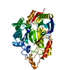
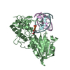
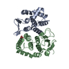
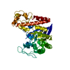
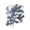
| Title | Crystal structure of APRT2 from Thermus thermophilus HB8 | ||||||
 Components Components | Adenine phosphoribosyltransferase | ||||||
 Keywords Keywords | TRANSFERASE / Phosphoribosyltransferase / Thermostable / Enzyme / Nucleic acid | ||||||
| Function / homology | Rossmann fold - #2020 / Phosphoribosyl transferase domain / Phosphoribosyltransferase-like / Phosphoribosyltransferase domain / glycosyltransferase activity / Rossmann fold / 3-Layer(aba) Sandwich / Alpha Beta / Adenine phosphoribosyltransferase Function and homology information Function and homology information | ||||||
| Biological species |   Thermus thermophilus (bacteria) Thermus thermophilus (bacteria) | ||||||
| Method |  X-RAY DIFFRACTION / SYNCHROTRON / MOLECULAR REPLACEMENT / Resolution: 2.6 Å X-RAY DIFFRACTION / SYNCHROTRON / MOLECULAR REPLACEMENT / Resolution: 2.6 Å | ||||||
 Authors Authors | Kunishima, N. / Naitow, H. / Matsuura, Y. | ||||||
 Citation Citation | Journal: Bioresour. Technol. / Year: 2019 Title: Structural and functional characterization of thermostable biocatalysts for the synthesis of 6-aminopurine nucleoside-5'-monophospate analogues. Authors: Arco, J.D. / Perez, E. / Naitow, H. / Matsuura, Y. / Kunishima, N. / Fernandez-Lucas, J. | ||||||
| History |
|
- Structure visualization
| Structure viewer | Molecule: MolmilJmol/JSmol |
|---|
- Downloads & links
-Download
| PDBx/mmCIF format | 5zgo.cif.gz | 205.4 KB | Display | PDBx/mmCIF format |
|---|---|---|---|---|
| PDB format | pdb5zgo.ent.gz | 166.8 KB | Display | PDB format |
| PDBx/mmJSON format | 5zgo.json.gz | Tree view | PDBx/mmJSON format | |
| Others |  Other downloads Other downloads |
-Validation report
| Arichive directory | https://data.pdbj.org/pub/pdb/validation_reports/zg/5zgoftp://data.pdbj.org/pub/pdb/validation_reports/zg/5zgo | HTTPS FTP |
|---|
-Related structure data
| Related structure data |  1vchS S: Starting model for refinement |
|---|---|
| Similar structure data |


-Links
 PDBj
PDBj
- Assembly
| Deposited unit | 
| ||||||||
|---|---|---|---|---|---|---|---|---|---|
| 1 | 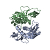
| ||||||||
| 2 | 
| ||||||||
| 3 | 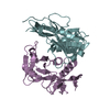
| ||||||||
| Unit cell |
|
-Components
| #1: Protein | Mass: 18912.135 Da / Num. of mol.: 6 Source method: isolated from a genetically manipulated source Source: (gene. exp.) Thermus thermophilus (strain HB8 / ATCC 27634 / DSM 579) (bacteria)Strain: HB8 / ATCC 27634 / DSM 579 / Gene: TTHA1614 / Production host: #2: Water | ChemComp-HOH / |  Mass: 18.015 Da / Num. of mol.: 155 / Source method: isolated from a natural source / Formula: H2O Mass: 18.015 Da / Num. of mol.: 155 / Source method: isolated from a natural source / Formula: H2O |
|---|
-Experimental details
-Experiment
| Experiment | Method: X-RAY DIFFRACTION / Number of used crystals: 1 |
|---|
- Sample preparation
| Crystal | Density Matthews: 2.3 Å3/Da / Density % sol: 46.57 % |
|---|---|
| Crystal grow | Temperature: 293 K / Method: vapor diffusion, hanging drop / pH: 6 / Details: 19% PEG 20K, 0.1 M Citrate-NaOH pH 6.0 |
-Data collection
| Diffraction | Mean temperature: 100 K |
|---|---|
| Diffraction source | Source: SYNCHROTRON / Site: SPring-8  / Beamline: BL26B2 / Wavelength: 1 Å / Beamline: BL26B2 / Wavelength: 1 Å |
| Detector | Type: MARMOSAIC 225 mm CCD / Detector: CCD / Date: Oct 5, 2017 |
| Radiation | Protocol: SINGLE WAVELENGTH / Monochromatic (M) / Laue (L): M / Scattering type: x-ray |
| Radiation wavelength | Wavelength: 1 Å / Relative weight: 1 |
| Reflection | Resolution: 2.6→45 Å / Num. obs: 31724 / % possible obs: 99.3 % / Redundancy: 3.6 % / Biso Wilson estimate: 46.17 Å2 / Rmerge(I) obs: 0.073 / Rpim(I) all: 0.044 / Rrim(I) all: 0.086 / Χ2: 1.611 / Net I/av σ(I): 24.64 / Net I/σ(I): 16.95 |
| Reflection shell | Resolution: 2.6→2.64 Å / Redundancy: 3.5 % / Rmerge(I) obs: 0.333 / Mean I/σ(I) obs: 4.03 / Num. unique obs: 1569 / CC1/2: 0.907 / Rpim(I) all: 0.204 / Rrim(I) all: 0.392 / Χ2: 0.868 / % possible all: 98.9 |
- Processing
| Software |
| ||||||||||||||||||||
|---|---|---|---|---|---|---|---|---|---|---|---|---|---|---|---|---|---|---|---|---|---|
| Refinement | Method to determine structure: MOLECULAR REPLACEMENT Starting model: 1VCH Resolution: 2.6→45 Å / Cross valid method: THROUGHOUT / Phase error: 30.03
| ||||||||||||||||||||
| Solvent computation | Shrinkage radii: 0.9 Å / VDW probe radii: 1.11 Å | ||||||||||||||||||||
| Displacement parameters | Biso mean: 47.39 Å2 | ||||||||||||||||||||
| Refinement step | Cycle: LAST / Resolution: 2.6→45 Å
| ||||||||||||||||||||
| LS refinement shell | Resolution: 2.6→2.68 Å
|
