 Movie
Movie Controller

[English] 日本語
 Yorodumi
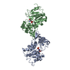
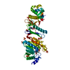
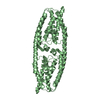
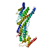
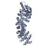
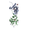
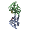
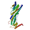
- PDB-5z98: Crystal Structure of the Primate APOBEC3H Dimer mediated by RNA Duplex -
+ Open data

- Basic information
| Entry | Database: PDB / ID: 5z98 | ||||||
|---|---|---|---|---|---|---|---|
| Title | Crystal Structure of the Primate APOBEC3H Dimer mediated by RNA Duplex | ||||||
 Components Components |
| ||||||
 Keywords Keywords | ANTIVIRAL PROTEIN/RNA / APOBEC3 / APOBEC3H / cytidine deaminase / deaminase / anti-HIV / Vif / dsRNA / cancer / HIV-1 / SIV / chimpanzee / dimerization / Pan troglodytes / ANTIVIRAL PROTEIN-RNA complex | ||||||
| Function / homology |  Function and homology information Function and homology informationsingle-stranded DNA cytosine deaminase / clearance of foreign intracellular DNA / cytidine deaminase activity / transposable element silencing / negative regulation of viral genome replication / defense response to virus / zinc ion binding / cytoplasm Similarity search - Function | ||||||
| Biological species |  unidentified (others) | ||||||
| Method |  X-RAY DIFFRACTION / SYNCHROTRON / MOLECULAR REPLACEMENT / Resolution: 2.2 Å X-RAY DIFFRACTION / SYNCHROTRON / MOLECULAR REPLACEMENT / Resolution: 2.2 Å | ||||||
 Authors Authors | Matsuoka, T. / Nagae, T. / Ode, H. / Watanabe, N. / Iwatani, Y. | ||||||
| Funding support |  Japan, 1items Japan, 1items
| ||||||
 Citation Citation | Journal: Nucleic Acids Res. / Year: 2018 Title: Structural basis of chimpanzee APOBEC3H dimerization stabilized by double-stranded RNA. Authors: Matsuoka, T. / Nagae, T. / Ode, H. / Awazu, H. / Kurosawa, T. / Hamano, A. / Matsuoka, K. / Hachiya, A. / Imahashi, M. / Yokomaku, Y. / Watanabe, N. / Iwatani, Y. | ||||||
| History |
|
- Structure visualization
| Structure viewer | Molecule: MolmilJmol/JSmol |
|---|
- Downloads & links
-Download
| PDBx/mmCIF format | 5z98.cif.gz | 102.2 KB | Display | PDBx/mmCIF format |
|---|---|---|---|---|
| PDB format | pdb5z98.ent.gz | 74.3 KB | Display | PDB format |
| PDBx/mmJSON format | 5z98.json.gz | Tree view | PDBx/mmJSON format | |
| Others |  Other downloads Other downloads |
-Validation report
| Arichive directory | https://data.pdbj.org/pub/pdb/validation_reports/z9/5z98ftp://data.pdbj.org/pub/pdb/validation_reports/z9/5z98 | HTTPS FTP |
|---|
-Related structure data
| Related structure data |  5w3vS S: Starting model for refinement |
|---|---|
| Similar structure data |


-Links
 PDBj
PDBj

- Assembly
| Deposited unit | 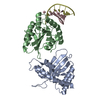
| ||||||||
|---|---|---|---|---|---|---|---|---|---|
| 1 | 
| ||||||||
| Unit cell |
|
-Components
| #1: Protein | Mass: 21912.309 Da / Num. of mol.: 2 Source method: isolated from a genetically manipulated source Source: (gene. exp.)  #2: RNA chain | | Mass: 3192.948 Da / Num. of mol.: 1 Source method: isolated from a genetically manipulated source Source: (gene. exp.) unidentified (others) / Production host: #3: RNA chain | | Mass: 3159.965 Da / Num. of mol.: 1 Source method: isolated from a genetically manipulated source Source: (gene. exp.) unidentified (others) / Production host: #4: Chemical |   Mass: 65.409 Da / Num. of mol.: 2 / Source method: obtained synthetically / Formula: Zn Mass: 65.409 Da / Num. of mol.: 2 / Source method: obtained synthetically / Formula: Zn#5: Water | ChemComp-HOH / |  Mass: 18.015 Da / Num. of mol.: 77 / Source method: isolated from a natural source / Formula: H2O Mass: 18.015 Da / Num. of mol.: 77 / Source method: isolated from a natural source / Formula: H2OHas protein modification | Y | |
|---|
-Experimental details
-Experiment
| Experiment | Method: X-RAY DIFFRACTION / Number of used crystals: 1 |
|---|
- Sample preparation
| Crystal | Density Matthews: 2.54 Å3/Da / Density % sol: 51.6 % |
|---|---|
| Crystal grow | Temperature: 293 K / Method: vapor diffusion, hanging drop / Details: 10% PEG 3350, 100mM ammonium formate |
-Data collection
| Diffraction | Mean temperature: 100 K |
|---|---|
| Diffraction source | Source: SYNCHROTRON / Site: SPring-8 / Beamline: BL41XU / Wavelength: 1 Å |
| Detector | Type: DECTRIS PILATUS3 S 6M / Detector: PIXEL / Date: Dec 7, 2017 |
| Radiation | Protocol: SINGLE WAVELENGTH / Monochromatic (M) / Laue (L): M / Scattering type: x-ray |
| Radiation wavelength | Wavelength: 1 Å / Relative weight: 1 |
| Reflection | Resolution: 2.2→43.49 Å / Num. obs: 24557 / % possible obs: 97.3 % / Redundancy: 3.1 % / CC1/2: 0.996 / Rmerge(I) obs: 0.073 / Net I/σ(I): 8.5 |
| Reflection shell | Resolution: 2.2→2.27 Å / Redundancy: 2.8 % / Rmerge(I) obs: 0.471 / Mean I/σ(I) obs: 2.1 / Num. unique obs: 2074 / CC1/2: 0.876 / % possible all: 94.7 |
- Processing
| Software |
| |||||||||||||||||||||||||||||||||||||||||||||||||||||||||||||||
|---|---|---|---|---|---|---|---|---|---|---|---|---|---|---|---|---|---|---|---|---|---|---|---|---|---|---|---|---|---|---|---|---|---|---|---|---|---|---|---|---|---|---|---|---|---|---|---|---|---|---|---|---|---|---|---|---|---|---|---|---|---|---|---|---|
| Refinement | Method to determine structure: MOLECULAR REPLACEMENT Starting model: 5w3v Resolution: 2.2→43.485 Å / SU ML: 0.34 / Cross valid method: NONE / σ(F): 1.34 / Phase error: 42.36
| |||||||||||||||||||||||||||||||||||||||||||||||||||||||||||||||
| Solvent computation | Shrinkage radii: 0.9 Å / VDW probe radii: 1.11 Å | |||||||||||||||||||||||||||||||||||||||||||||||||||||||||||||||
| Refinement step | Cycle: LAST / Resolution: 2.2→43.485 Å
| |||||||||||||||||||||||||||||||||||||||||||||||||||||||||||||||
| Refine LS restraints |
| |||||||||||||||||||||||||||||||||||||||||||||||||||||||||||||||
| LS refinement shell |
|
