 Movie
Movie Controller

[English] 日本語
 Yorodumi
- PDB-5z6c: Crystal structure of Bacillus subtilis sugar-binding protein YesO... -
+ Open data

- Basic information
| Entry | Database: PDB / ID: 5z6c | ||||||
|---|---|---|---|---|---|---|---|
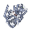
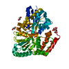
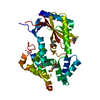
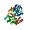
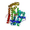
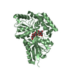
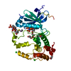
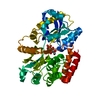
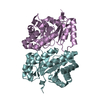
| Title | Crystal structure of Bacillus subtilis sugar-binding protein YesO involved in import of rhamnogalacturonan | ||||||
 Components Components | Putative ABC transporter substrate-binding protein YesO | ||||||
 Keywords Keywords | TRANSPORT PROTEIN / ABC transporter substrate-binding protein / sugar transport | ||||||
| Function / homology |  Function and homology information Function and homology information | ||||||
| Biological species |  | ||||||
| Method |  X-RAY DIFFRACTION / SYNCHROTRON / MOLECULAR REPLACEMENT / Resolution: 1.97 Å X-RAY DIFFRACTION / SYNCHROTRON / MOLECULAR REPLACEMENT / Resolution: 1.97 Å | ||||||
 Authors Authors | Sugiura, H. / Oiki, S. / Mikami, B. / Murata, K. / Hashimoto, W. | ||||||
| Funding support |  Japan, 1items Japan, 1items
| ||||||
 Citation Citation | Journal: To Be Published Title: Crystal structure of Bacillus subtilis sugar-binding protein YesO involved in import of rhamnogalacturonan Authors: Sugiura, H. / Oiki, S. / Mikami, B. / Murata, K. / Hashimoto, W. | ||||||
| History |
|
- Structure visualization
| Structure viewer | Molecule: MolmilJmol/JSmol |
|---|
- Downloads & links
-Download
| PDBx/mmCIF format | 5z6c.cif.gz | 91.7 KB | Display | PDBx/mmCIF format |
|---|---|---|---|---|
| PDB format | pdb5z6c.ent.gz | 66.7 KB | Display | PDB format |
| PDBx/mmJSON format | 5z6c.json.gz | Tree view | PDBx/mmJSON format | |
| Others |  Other downloads Other downloads |
-Validation report
| Arichive directory | https://data.pdbj.org/pub/pdb/validation_reports/z6/5z6cftp://data.pdbj.org/pub/pdb/validation_reports/z6/5z6c | HTTPS FTP |
|---|
-Related structure data
| Related structure data |  4r6kS S: Starting model for refinement |
|---|---|
| Similar structure data |

-Links
 PDBj
PDBj


- Assembly
| Deposited unit | 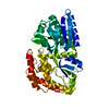
| ||||||||
|---|---|---|---|---|---|---|---|---|---|
| 1 |
| ||||||||
| Unit cell |
|
-Components
| #1: Protein | Mass: 48390.648 Da / Num. of mol.: 1 Source method: isolated from a genetically manipulated source Source: (gene. exp.) Strain: 168 / Gene: yesO, BSU06970 / Production host: |
|---|---|
| #2: Water | ChemComp-HOH /  Mass: 18.015 Da / Num. of mol.: 116 / Source method: isolated from a natural source / Formula: H2O Mass: 18.015 Da / Num. of mol.: 116 / Source method: isolated from a natural source / Formula: H2O |
-Experimental details
-Experiment
| Experiment | Method: X-RAY DIFFRACTION / Number of used crystals: 1 |
|---|
- Sample preparation
| Crystal | Density Matthews: 1.61 Å3/Da / Density % sol: 23.62 % |
|---|---|
| Crystal grow | Temperature: 293 K / Method: vapor diffusion, sitting drop Details: 0.1M MIB (sodium malonate, imidazole, boric acid) buffer (pH 6.0), 25% (w/v) PEG-1500 |
-Data collection
| Diffraction | Mean temperature: 100 K |
|---|---|
| Diffraction source | Source: SYNCHROTRON / Site: SPring-8 / Beamline: BL38B1 / Wavelength: 1 Å |
| Detector | Type: MARMOSAIC 225 mm CCD / Detector: CCD / Date: Apr 22, 2016 |
| Radiation | Protocol: SINGLE WAVELENGTH / Monochromatic (M) / Laue (L): M / Scattering type: x-ray |
| Radiation wavelength | Wavelength: 1 Å / Relative weight: 1 |
| Reflection | Resolution: 1.97→50 Å / Num. obs: 42265 / % possible obs: 98.9 % / Redundancy: 2.2 % / Rmerge(I) obs: 0.12 / Net I/σ(I): 12.1 |
| Reflection shell | Resolution: 1.97→2 Å / Redundancy: 2.2 % / Rmerge(I) obs: 0.278 / Mean I/σ(I) obs: 3.06 / % possible all: 98.3 |
- Processing
| Software |
| |||||||||||||||||||||||||||||||||||||||||||||||||||||||||||||||
|---|---|---|---|---|---|---|---|---|---|---|---|---|---|---|---|---|---|---|---|---|---|---|---|---|---|---|---|---|---|---|---|---|---|---|---|---|---|---|---|---|---|---|---|---|---|---|---|---|---|---|---|---|---|---|---|---|---|---|---|---|---|---|---|---|
| Refinement | Method to determine structure: MOLECULAR REPLACEMENT Starting model: 4R6K Resolution: 1.97→37.261 Å / SU ML: 0.22 / Cross valid method: FREE R-VALUE / σ(F): 1.42 / Phase error: 26.89
| |||||||||||||||||||||||||||||||||||||||||||||||||||||||||||||||
| Solvent computation | Shrinkage radii: 0.9 Å / VDW probe radii: 1.11 Å | |||||||||||||||||||||||||||||||||||||||||||||||||||||||||||||||
| Refinement step | Cycle: LAST / Resolution: 1.97→37.261 Å
| |||||||||||||||||||||||||||||||||||||||||||||||||||||||||||||||
| Refine LS restraints |
| |||||||||||||||||||||||||||||||||||||||||||||||||||||||||||||||
| LS refinement shell |
|
