| Entry | Database: PDB / ID: 5z3m
|
|---|
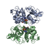
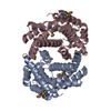
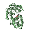
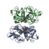
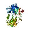
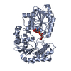
| Title | Crystal structure of Low Molecular Weight Phosphotyrosine phosphatase (VcLMWPTP-2) from Vibrio choleraeO395 |
|---|
 Components Components | Phosphotyrosine protein phosphatase |
|---|
 Keywords Keywords | HYDROLASE / Phosphatase / Ligand Bound |
|---|
| Function / homology |  Function and homology information Function and homology information
: / Protein-tyrosine phosphatase, low molecular weight / Phosphotyrosine protein phosphatase I / Phosphotyrosine protein phosphatase I superfamily / Low molecular weight phosphotyrosine protein phosphatase / Low molecular weight phosphatase family / Response regulator / Rossmann fold / 3-Layer(aba) Sandwich / Alpha BetaSimilarity search - Domain/homology |
|---|
| Biological species |  Vibrio cholerae serotype O1 (bacteria) Vibrio cholerae serotype O1 (bacteria) |
|---|
| Method |  X-RAY DIFFRACTION / SYNCHROTRON / MOLECULAR REPLACEMENT / Resolution: 2.6 Å X-RAY DIFFRACTION / SYNCHROTRON / MOLECULAR REPLACEMENT / Resolution: 2.6 Å |
|---|
 Authors Authors | Chatterjee, S. / Nath, S. / Sen, U. |
|---|
 Citation Citation | Journal: Biochim Biophys Acta Proteins Proteom / Year: 2019
Title: Vibrio cholerae LMWPTP-2 display unique surface charge and grooves around the active site: Indicative of distinctive substrate specificity and scope to design specific inhibitor.
Authors: Chatterjee, S. / Nath, S. / Ghosh, B. / Sen, U. |
|---|
| History | | Deposition | Jan 8, 2018 | Deposition site: PDBJ / Processing site: PDBJ |
|---|
| Revision 1.0 | Sep 19, 2018 | Provider: repository / Type: Initial release |
|---|
| Revision 1.1 | May 13, 2020 | Group: Database references / Category: citation / citation_author
Item: _citation.journal_abbrev / _citation.journal_id_CSD ..._citation.journal_abbrev / _citation.journal_id_CSD / _citation.journal_id_ISSN / _citation.journal_volume / _citation.page_first / _citation.page_last / _citation.pdbx_database_id_DOI / _citation.pdbx_database_id_PubMed / _citation.title / _citation.year |
|---|
| Revision 1.2 | Nov 22, 2023 | Group: Data collection / Database references / Refinement description
Category: chem_comp_atom / chem_comp_bond ...chem_comp_atom / chem_comp_bond / database_2 / pdbx_initial_refinement_model
Item: _database_2.pdbx_DOI / _database_2.pdbx_database_accession |
|---|
|
|---|
 Movie
Movie Controller

 Yorodumi
 Open data
 Basic information
 Structure visualization
 Downloads & links
 Other downloads


 PDBj
PDBj
 Assembly
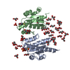

 Mass: 209.263 Da / Num. of mol.: 2 / Source method: obtained synthetically / Formula: C7H15NO4S / Comment: pH buffer*YM
 Mass: 96.063 Da / Num. of mol.: 2 / Source method: obtained synthetically / Formula: SO4
 Mass: 92.094 Da / Num. of mol.: 35 / Source method: obtained synthetically / Formula: C3H8O3
 Mass: 18.015 Da / Num. of mol.: 256 / Source method: isolated from a natural source / Formula: H2O
 Sample preparation
 / Beamline: PX-BL21 / Wavelength: 0.98 Å
 Processing