[English] 日本語
Yorodumi
- PDB-5z10: Structure of the mechanosensitive Piezo1 channel -

+
Open data


ID or keywords:

Loading...

-
Basic information

Entry
Database: PDB / ID: 5z10
TitleStructure of the mechanosensitive Piezo1 channel
ComponentsPiezo-type mechanosensitive ion channel component 1
KeywordsMEMBRANE PROTEIN / Piezo1 / Piezo2 / mechanogating / channel
Function / homology
Function and homology information


mechanosensitive monoatomic cation channel activity / positive regulation of cell-cell adhesion mediated by integrin / detection of mechanical stimulus / mechanosensitive monoatomic ion channel activity / positive regulation of integrin activation / positive regulation of myotube differentiation / lamellipodium membrane / monoatomic cation transport / monoatomic cation channel activity / regulation of membrane potential ...mechanosensitive monoatomic cation channel activity / positive regulation of cell-cell adhesion mediated by integrin / detection of mechanical stimulus / mechanosensitive monoatomic ion channel activity / positive regulation of integrin activation / positive regulation of myotube differentiation / lamellipodium membrane / monoatomic cation transport / monoatomic cation channel activity / regulation of membrane potential / endoplasmic reticulum-Golgi intermediate compartment membrane / cellular response to mechanical stimulus / endoplasmic reticulum membrane / endoplasmic reticulum / identical protein binding / plasma membrane
Similarity search - Function
Piezo family / Piezo non-specific cation channel, R-Ras-binding domain / Piezo domain / Piezo non-specific cation channel, R-Ras-binding domain / Piezo
Similarity search - Domain/homology
Piezo-type mechanosensitive ion channel component 1
Similarity search - Component
Biological speciesMus musculus (house mouse)
MethodELECTRON MICROSCOPY / single particle reconstruction / cryo EM / Resolution: 3.97 Å
AuthorsZhao, Q. / Zhou, H. / Chi, S. / Wang, Y. / Wang, J. / Geng, J. / Wu, K. / Liu, W. / Zhang, T. / Dong, M.-Q. ...Zhao, Q. / Zhou, H. / Chi, S. / Wang, Y. / Wang, J. / Geng, J. / Wu, K. / Liu, W. / Zhang, T. / Dong, M.-Q. / Wang, J. / Li, X. / Xiao, B.
Funding support China, 1items
OrganizationGrant numberCountry
National Natural Science Foundation of China (NSFC)31630090 China
Citation
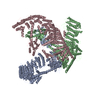
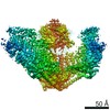
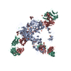
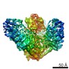
Journal: Nature / Year: 2018
Title: Structure and mechanogating mechanism of the Piezo1 channel.
Authors: Qiancheng Zhao / Heng Zhou / Shaopeng Chi / Yanfeng Wang / Jianhua Wang / Jie Geng / Kun Wu / Wenhao Liu / Tingxin Zhang / Meng-Qiu Dong / Jiawei Wang / Xueming Li / Bailong Xiao /
Abstract: The mechanosensitive Piezo channels function as key eukaryotic mechanotransducers. However, their structures and mechanogating mechanisms remain unknown. Here we determine the three-bladed, propeller- ...The mechanosensitive Piezo channels function as key eukaryotic mechanotransducers. However, their structures and mechanogating mechanisms remain unknown. Here we determine the three-bladed, propeller-like electron cryo-microscopy structure of mouse Piezo1 and functionally reveal its mechanotransduction components. Despite the lack of sequence repetition, we identify nine repetitive units consisting of four transmembrane helices each-which we term transmembrane helical units (THUs)-which assemble into a highly curved blade-like structure. The last transmembrane helix encloses a hydrophobic pore, followed by three intracellular fenestration sites and side portals that contain pore-property-determining residues. The central region forms a 90 Å-long intracellular beam-like structure, which undergoes a lever-like motion to connect THUs to the pore via the interfaces of the C-terminal domain, the anchor-resembling domain and the outer helix. Deleting extracellular loops in the distal THUs or mutating single residues in the beam impairs the mechanical activation of Piezo1. Overall, Piezo1 possesses a unique 38-transmembrane-helix topology and designated mechanotransduction components, which enable a lever-like mechanogating mechanism.
History
DepositionDec 22, 2017Deposition site: PDBJ / Processing site: PDBJ
Revision 1.0Jan 31, 2018Provider: repository / Type: Initial release
Revision 1.1Feb 7, 2018Group: Database references / Structure summary / Category: audit_author / citation_author / Item: _audit_author.name / _citation_author.name
Revision 1.2Mar 7, 2018Group: Database references / Category: citation / citation_author
Item: _citation.journal_volume / _citation.page_first ..._citation.journal_volume / _citation.page_first / _citation.page_last / _citation.pdbx_database_id_PubMed / _citation.title / _citation_author.name
Revision 2.0Jan 29, 2020Group: Advisory / Atomic model ...Advisory / Atomic model / Author supporting evidence / Data collection / Derived calculations / Refinement description / Structure summary
Category: atom_site / computing ...atom_site / computing / em_software / pdbx_audit_support / pdbx_poly_seq_scheme / pdbx_struct_assembly_prop / pdbx_struct_sheet_hbond / pdbx_unobs_or_zero_occ_atoms / pdbx_unobs_or_zero_occ_residues / pdbx_validate_peptide_omega / pdbx_validate_rmsd_angle / pdbx_validate_torsion / refine_ls_restr / software / struct_conf / struct_conn / struct_sheet / struct_sheet_order / struct_sheet_range
Item: _em_software.category / _pdbx_poly_seq_scheme.auth_mon_id ..._em_software.category / _pdbx_poly_seq_scheme.auth_mon_id / _pdbx_poly_seq_scheme.auth_seq_num / _pdbx_poly_seq_scheme.pdb_mon_id / _refine_ls_restr.dev_ideal / _refine_ls_restr.number / _software.version / _struct_conn.pdbx_dist_value
Description: Model completeness / Provider: author / Type: Coordinate replacement

-
Structure visualization

Movie
  • Deposited structure unit
  • Imaged by Jmol
  • Download
  • Superimposition on EM map
  • EMDB-6865
  • Imaged by UCSF Chimera
  • Download
Movie viewer
Structure viewerMolecule:
MolmilJmol/JSmol

Downloads & links

-
Assembly

Deposited unit
A: Piezo-type mechanosensitive ion channel component 1
B: Piezo-type mechanosensitive ion channel component 1
C: Piezo-type mechanosensitive ion channel component 1


Theoretical massNumber of molelcules
Total (without water)876,9623
Polymers876,9623
Non-polymers00
Water0
1


  • Idetical with deposited unit
  • defined by author
  • Evidence: gel filtration
TypeNameSymmetry operationNumber
identity operation1_5551

-
Components

#1: Protein Piezo-type mechanosensitive ion channel component 1 / Protein FAM38A


Mass: 292320.656 Da / Num. of mol.: 3
Source method: isolated from a genetically manipulated source
Source: (gene. exp.) Mus musculus (house mouse) / Gene: Piezo1, Fam38a / Cell line (production host): HEK293T / Production host: 
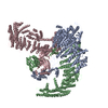
Homo sapiens (human) / References: UniProt: E2JF22

-
Experimental details

-
Experiment

ExperimentMethod: ELECTRON MICROSCOPY
EM experimentAggregation state: PARTICLE / 3D reconstruction method: single particle reconstruction

-
Sample preparation

ComponentName: Cryo-EM map of the mechanosensitive Piezo1 channel / Type: COMPLEX / Entity ID: all / Source: RECOMBINANT
Source (natural)Organism: Mus musculus (house mouse)
Source (recombinant)Organism: Homo sapiens (human) / Cell: HEK293T
Buffer solutionpH: 7.2
SpecimenConc.: 0.18 mg/ml / Embedding applied: NO / Shadowing applied: NO / Staining applied: NO / Vitrification applied: YES
Specimen supportGrid material: COPPER / Grid mesh size: 300 divisions/in. / Grid type: Quantifoil R2/2
VitrificationInstrument: FEI VITROBOT MARK IV / Cryogen name: ETHANE / Humidity: 100 % / Chamber temperature: 281 K
Details: After a 15 sec waiting time, the grids were blotted for 3.5 sec and plunged into liquid ethane

-
Electron microscopy imaging

Experimental equipment
Model: Titan Krios / Image courtesy: FEI Company
MicroscopyModel: FEI TITAN KRIOS
Electron gunElectron source: FIELD EMISSION GUN / Accelerating voltage: 300 kV / Illumination mode: FLOOD BEAM
Electron lensMode: BRIGHT FIELDBright-field microscopy / Nominal magnification: 22500 X / Nominal defocus min: 1500 nm / Calibrated defocus min: 3000 nm / Cs: 2.7 mm / C2 aperture diameter: 50 µm / Alignment procedure: COMA FREE
Specimen holderCryogen: NITROGEN / Specimen holder model: FEI TITAN KRIOS AUTOGRID HOLDER
Image recordingAverage exposure time: 8 sec. / Electron dose: 50 e/Å2 / Detector mode: SUPER-RESOLUTION / Film or detector model: GATAN K2 SUMMIT (4k x 4k)
Image scansMovie frames/image: 32 / Used frames/image: 1-32

-
Processing

SoftwareName: PHENIX / Version: dev_3374: / Classification: refinement
EM software
IDNameVersionCategory
2UCSFImage4image acquisition
4RELION1.4CTF correction
10RELION1.4initial Euler assignment
11RELION1.4final Euler assignment
12RELION1.4classification
13RELION1.43D reconstruction
CTF correctionType: PHASE FLIPPING AND AMPLITUDE CORRECTION
SymmetryPoint symmetry: C3 (3 fold cyclic)
3D reconstructionResolution: 3.97 Å / Resolution method: FSC 0.143 CUT-OFF / Num. of particles: 238529 / Symmetry type: POINT
Refine LS restraints
Refine-IDTypeDev idealNumber
ELECTRON MICROSCOPYf_bond_d0.00629763
ELECTRON MICROSCOPYf_angle_d1.02940506
ELECTRON MICROSCOPYf_dihedral_angle_d10.10717556
ELECTRON MICROSCOPYf_chiral_restr0.0594716
ELECTRON MICROSCOPYf_plane_restr0.0085148

+
About Yorodumi

-
News

-
Feb 9, 2022. New format data for meta-information of EMDB entries

New format data for meta-information of EMDB entries

  • Version 3 of the EMDB header file is now the official format.
  • The previous official version 1.9 will be removed from the archive.

Related info.:EMDB header

External links:wwPDB to switch to version 3 of the EMDB data model

-
Aug 12, 2020. Covid-19 info

Covid-19 info

URL: https://pdbj.org/emnavi/covid19.php

New page: Covid-19 featured information page in EM Navigator.

Related info.:Covid-19 info / Mar 5, 2020. Novel coronavirus structure data

+
Mar 5, 2020. Novel coronavirus structure data

Novel coronavirus structure data

Related info.:Yorodumi Speices / Aug 12, 2020. Covid-19 info

External links:COVID-19 featured content - PDBj / Molecule of the Month (242):Coronavirus Proteases

+
Jan 31, 2019. EMDB accession codes are about to change! (news from PDBe EMDB page)

EMDB accession codes are about to change! (news from PDBe EMDB page)

  • The allocation of 4 digits for EMDB accession codes will soon come to an end. Whilst these codes will remain in use, new EMDB accession codes will include an additional digit and will expand incrementally as the available range of codes is exhausted. The current 4-digit format prefixed with “EMD-” (i.e. EMD-XXXX) will advance to a 5-digit format (i.e. EMD-XXXXX), and so on. It is currently estimated that the 4-digit codes will be depleted around Spring 2019, at which point the 5-digit format will come into force.
  • The EM Navigator/Yorodumi systems omit the EMD- prefix.

Related info.:Q: What is EMD? / ID/Accession-code notation in Yorodumi/EM Navigator

External links:EMDB Accession Codes are Changing Soon! / Contact to PDBj

+
Jul 12, 2017. Major update of PDB

Major update of PDB

  • wwPDB released updated PDB data conforming to the new PDBx/mmCIF dictionary.
  • This is a major update changing the version number from 4 to 5, and with Remediation, in which all the entries are updated.
  • In this update, many items about electron microscopy experimental information are reorganized (e.g. em_software).
  • Now, EM Navigator and Yorodumi are based on the updated data.

External links:wwPDB Remediation / Enriched Model Files Conforming to OneDep Data Standards Now Available in the PDB FTP Archive

-
Yorodumi

Thousand views of thousand structures

  • Yorodumi is a browser for structure data from EMDB, PDB, SASBDB, etc.
  • This page is also the successor to EM Navigator detail page, and also detail information page/front-end page for Omokage search.
  • The word "yorodu" (or yorozu) is an old Japanese word meaning "ten thousand". "mi" (miru) is to see.

Related info.:EMDB / PDB / SASBDB / Comparison of 3 databanks / Yorodumi Search / Aug 31, 2016. New EM Navigator & Yorodumi / Yorodumi Papers / Jmol/JSmol / Function and homology information / Changes in new EM Navigator and Yorodumi

Read more