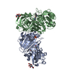
- PDB-5yt3: Structure of the Human Mitogen-Activated Protein Kinase Kinase 1 ... -
+
Open data
ID or keywords:
Loading...
-
Basic information
Entry
Database: PDB / ID: 5yt3
Title
Structure of the Human Mitogen-Activated Protein Kinase Kinase 1 S218D and S222D mutant
Components
Mitogen-activated protein kinase kinase 1, isoform CRA_d
Keywords
TRANSFERASE / Kinase
Function / homology
Function and homology information
epithelial cell proliferation involved in lung morphogenesis / negative regulation of homotypic cell-cell adhesion / positive regulation of endodermal cell differentiation / negative regulation of hypoxia-induced intrinsic apoptotic signaling pathway / regulation of vascular associated smooth muscle contraction / regulation of axon regeneration / mitogen-activated protein kinase kinase / positive regulation of muscle contraction / placenta blood vessel development / Golgi inheritance ...epithelial cell proliferation involved in lung morphogenesis / negative regulation of homotypic cell-cell adhesion / positive regulation of endodermal cell differentiation / negative regulation of hypoxia-induced intrinsic apoptotic signaling pathway / regulation of vascular associated smooth muscle contraction / regulation of axon regeneration / mitogen-activated protein kinase kinase / positive regulation of muscle contraction / placenta blood vessel development / Golgi inheritance / MAP kinase scaffold activity / cerebellar cortex formation / labyrinthine layer development / melanosome transport / Signaling by MAP2K mutants / type B pancreatic cell proliferation / central nervous system neuron differentiation / vesicle transport along microtubule / positive regulation of Ras protein signal transduction / regulation of Golgi inheritance / mitogen-activated protein kinase kinase kinase binding / positive regulation of axonogenesis / trachea formation / triglyceride homeostasis / regulation of early endosome to late endosome transport / Negative feedback regulation of MAPK pathway / regulation of stress-activated MAPK cascade / Frs2-mediated activation / MAPK3 (ERK1) activation / ERBB2-ERBB3 signaling pathway / face development / MAP kinase kinase activity / endodermal cell differentiation / regulation of neurotransmitter receptor localization to postsynaptic specialization membrane / Bergmann glial cell differentiation / positive regulation of protein serine/threonine kinase activity / thyroid gland development / positive regulation of ATP biosynthetic process / Uptake and function of anthrax toxins / protein kinase activator activity / response to axon injury / Schwann cell development / keratinocyte differentiation / ERK1 and ERK2 cascade / neuron projection morphogenesis / myelination / protein serine/threonine/tyrosine kinase activity / insulin-like growth factor receptor signaling pathway / positive regulation of autophagy / dendrite cytoplasm / response to glucocorticoid / thymus development / Signal transduction by L1 / protein serine/threonine kinase activator activity / MAP3K8 (TPL2)-dependent MAPK1/3 activation / cell motility / positive regulation of transcription elongation by RNA polymerase II / RAF activation / Signaling by high-kinase activity BRAF mutants / MAP2K and MAPK activation / small GTPase binding / chemotaxis / neuron differentiation / cellular senescence / Signaling by RAF1 mutants / Signaling by moderate kinase activity BRAF mutants / Paradoxical activation of RAF signaling by kinase inactive BRAF / Signaling downstream of RAS mutants / Signaling by BRAF and RAF1 fusions / late endosome / MAPK cascade / heart development / protein tyrosine kinase activity / response to oxidative stress / scaffold protein binding / cell cortex / microtubule / perikaryon / early endosome / protein kinase activity / positive regulation of ERK1 and ERK2 cascade / postsynaptic density / positive regulation of cell migration / ciliary basal body / negative regulation of cell population proliferation / negative regulation of gene expression / protein serine kinase activity / axon / focal adhesion / protein serine/threonine kinase activity / positive regulation of gene expression / centrosome / positive regulation of DNA-templated transcription / protein-containing complex binding / perinuclear region of cytoplasm / glutamatergic synapse / Golgi apparatus / endoplasmic reticulum / signal transduction / mitochondrion Similarity search - Function
: / Transferase(Phosphotransferase) domain 1 / Transferase(Phosphotransferase); domain 1 / Serine/threonine-protein kinase, active site / Serine/Threonine protein kinases active-site signature. / Protein kinase domain / Serine/Threonine protein kinases, catalytic domain / Protein kinase, ATP binding site / Protein kinases ATP-binding region signature. / Protein kinase domain profile. ...: / Transferase(Phosphotransferase) domain 1 / Transferase(Phosphotransferase); domain 1 / Serine/threonine-protein kinase, active site / Serine/Threonine protein kinases active-site signature. / Protein kinase domain / Serine/Threonine protein kinases, catalytic domain / Protein kinase, ATP binding site / Protein kinases ATP-binding region signature. / Protein kinase domain profile. / Protein kinase domain / Protein kinase-like domain superfamily / Orthogonal Bundle / Mainly Alpha Similarity search - Domain/homology
 Movie
Movie Controller

 Yorodumi
 Open data
 Basic information
 Components
 Keywords
 Function and homology information
 Homo sapiens (human)
 X-RAY DIFFRACTION /
  Authors
 Citation
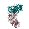
 Structure visualization
 Downloads & links
 Other downloads
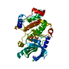


 PDBj
PDBj


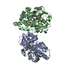
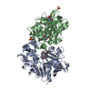
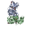
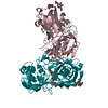
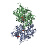
 Assembly
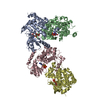


 Mass: 506.196 Da / Num. of mol.: 4 / Source method: obtained synthetically / Formula: C10H17N6O12P3 / Comment: AMP-PNP, energy-carrying molecule analogue*YM
 Mass: 24.305 Da / Num. of mol.: 2 / Source method: obtained synthetically / Formula: Mg
 Mass: 18.015 Da / Num. of mol.: 14 / Source method: isolated from a natural source / Formula: H2O
 Sample preparation
 / Beamline: BL38B1 / Wavelength: 1 Å
 Processing