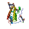
 Movie
Movie Controller


+ Open data

- Basic information
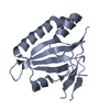
| Entry | Database: PDB / ID: 5ysi | ||||||
|---|---|---|---|---|---|---|---|
| Title | SdeA mART-C domain EE/AA NCA complex | ||||||
 Components Components | Ubiquitinating/deubiquitinating enzyme SdeA | ||||||
 Keywords Keywords | HYDROLASE / SdeA / E3 ligase / Legionella | ||||||
| Function / homology |  Function and homology information Function and homology informationNAD+-protein-arginine ADP-ribosyltransferase / NAD+-protein-arginine ADP-ribosyltransferase activity / deNEDDylase activity / protein deneddylation / Transferases; Acyltransferases; Aminoacyltransferases / K63-linked deubiquitinase activity / protein deubiquitination / nucleotidyltransferase activity / cysteine-type peptidase activity / host cell ...NAD+-protein-arginine ADP-ribosyltransferase / NAD+-protein-arginine ADP-ribosyltransferase activity / deNEDDylase activity / protein deneddylation / Transferases; Acyltransferases; Aminoacyltransferases / K63-linked deubiquitinase activity / protein deubiquitination / nucleotidyltransferase activity / cysteine-type peptidase activity / host cell / Hydrolases; Acting on peptide bonds (peptidases); Cysteine endopeptidases / protein ubiquitination / nucleotide binding / proteolysis / extracellular region Similarity search - Function | ||||||
| Biological species |  Legionella pneumophila subsp. pneumophila str. Philadelphia 1 (bacteria) Legionella pneumophila subsp. pneumophila str. Philadelphia 1 (bacteria) | ||||||
| Method |  X-RAY DIFFRACTION / SYNCHROTRON / SAD / Resolution: 1.546 Å X-RAY DIFFRACTION / SYNCHROTRON / SAD / Resolution: 1.546 Å | ||||||
 Authors Authors | Kim, L. / Kwon, D.H. / Song, H.K. | ||||||
 Citation Citation | Journal: J. Mol. Biol. / Year: 2018 Title: Structural and Biochemical Study of the Mono-ADP-Ribosyltransferase Domain of SdeA, a Ubiquitylating/Deubiquitylating Enzyme from Legionella pneumophila Authors: Kim, L. / Kwon, D.H. / Kim, B.H. / Kim, J. / Park, M.R. / Park, Z.Y. / Song, H.K. | ||||||
| History |
|
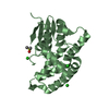
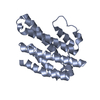
- Structure visualization
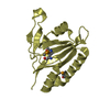
| Structure viewer | Molecule: MolmilJmol/JSmol |
|---|
- Downloads & links
-Download
| PDBx/mmCIF format | 5ysi.cif.gz | 68 KB | Display | PDBx/mmCIF format |
|---|---|---|---|---|
| PDB format | pdb5ysi.ent.gz | 50.4 KB | Display | PDB format |
| PDBx/mmJSON format | 5ysi.json.gz | Tree view | PDBx/mmJSON format | |
| Others |  Other downloads Other downloads |
-Validation report
| Arichive directory | https://data.pdbj.org/pub/pdb/validation_reports/ys/5ysiftp://data.pdbj.org/pub/pdb/validation_reports/ys/5ysi | HTTPS FTP |
|---|
-Related structure data


-Links
 PDBj
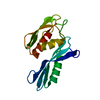
PDBj- Assembly
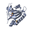
| Deposited unit | 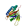
| ||||||||
|---|---|---|---|---|---|---|---|---|---|
| 1 |
| ||||||||
| Unit cell |
|
-Components
| #1: Protein | Mass: 16923.611 Da / Num. of mol.: 1 / Fragment: UNP residues 761-960 / Mutation: E860A, E862A Source method: isolated from a genetically manipulated source Source: (gene. exp.) Legionella pneumophila subsp. pneumophila str. Philadelphia 1 (bacteria)Strain: Philadelphia 1 / Gene: sdeA / Production host: References: UniProt: Q6RCR0, UniProt: Q5ZTK4*PLUS, Hydrolases; Acting on peptide bonds (peptidases); Cysteine endopeptidases, Transferases; Acyltransferases; Aminoacyltransferases, NAD+-protein- ...References: UniProt: Q6RCR0, UniProt: Q5ZTK4*PLUS, Hydrolases; Acting on peptide bonds (peptidases); Cysteine endopeptidases, Transferases; Acyltransferases; Aminoacyltransferases, NAD+-protein-arginine ADP-ribosyltransferase |
|---|---|
| #2: Chemical | ChemComp-NCA / 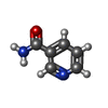  Mass: 122.125 Da / Num. of mol.: 1 / Source method: obtained synthetically / Formula: C6H6N2O / Comment: medication*YM Mass: 122.125 Da / Num. of mol.: 1 / Source method: obtained synthetically / Formula: C6H6N2O / Comment: medication*YM |
| #3: Water | ChemComp-HOH /  Mass: 18.015 Da / Num. of mol.: 32 / Source method: isolated from a natural source / Formula: H2O Mass: 18.015 Da / Num. of mol.: 32 / Source method: isolated from a natural source / Formula: H2O |
| Has protein modification | Y |
-Experimental details
-Experiment
| Experiment | Method: X-RAY DIFFRACTION / Number of used crystals: 1 |
|---|
- Sample preparation
| Crystal | Density Matthews: 1.78 Å3/Da / Density % sol: 30.93 % Description: THE ENTRY CONTAINS FRIEDEL PAIRS IN F_PLUS/MINUS COLUMNS. |
|---|---|
| Crystal grow | Temperature: 293 K / Method: vapor diffusion, sitting drop / Details: PEG400, Tris |
-Data collection
| Diffraction | Mean temperature: 193 K |
|---|---|
| Diffraction source | Source: SYNCHROTRON / Site: Photon Factory  / Beamline: BL-17A / Wavelength: 1 Å / Beamline: BL-17A / Wavelength: 1 Å |
| Detector | Type: ADSC QUANTUM 270 / Detector: CCD / Date: May 5, 2017 |
| Radiation | Protocol: SINGLE WAVELENGTH / Monochromatic (M) / Laue (L): M / Scattering type: x-ray |
| Radiation wavelength | Wavelength: 1 Å / Relative weight: 1 |
| Reflection | Resolution: 1.546→34.555 Å / Num. obs: 36284 / % possible obs: 99.56 % / Redundancy: 6.7 % / Net I/σ(I): 32.37 |
| Reflection shell | Resolution: 1.546→1.602 Å / Redundancy: 5.8 % / Rmerge(I) obs: 0.542 / Num. unique obs: 1754 / % possible all: 97.33 |
- Processing
| Software |
| |||||||||||||||||||||||||||||||||||||||||||||||||||||||||||||||||||||||||||||||||||||||||||||||||||||||||||||||||||||||||||||||||||||||||||||||||||||||||||||||||||||||||||||||
|---|---|---|---|---|---|---|---|---|---|---|---|---|---|---|---|---|---|---|---|---|---|---|---|---|---|---|---|---|---|---|---|---|---|---|---|---|---|---|---|---|---|---|---|---|---|---|---|---|---|---|---|---|---|---|---|---|---|---|---|---|---|---|---|---|---|---|---|---|---|---|---|---|---|---|---|---|---|---|---|---|---|---|---|---|---|---|---|---|---|---|---|---|---|---|---|---|---|---|---|---|---|---|---|---|---|---|---|---|---|---|---|---|---|---|---|---|---|---|---|---|---|---|---|---|---|---|---|---|---|---|---|---|---|---|---|---|---|---|---|---|---|---|---|---|---|---|---|---|---|---|---|---|---|---|---|---|---|---|---|---|---|---|---|---|---|---|---|---|---|---|---|---|---|---|---|---|
| Refinement | Method to determine structure: SAD / Resolution: 1.546→34.555 Å / SU ML: 0.2 / Cross valid method: FREE R-VALUE / σ(F): 1.37 / Phase error: 24.65
| |||||||||||||||||||||||||||||||||||||||||||||||||||||||||||||||||||||||||||||||||||||||||||||||||||||||||||||||||||||||||||||||||||||||||||||||||||||||||||||||||||||||||||||||
| Solvent computation | Shrinkage radii: 0.9 Å / VDW probe radii: 1.11 Å | |||||||||||||||||||||||||||||||||||||||||||||||||||||||||||||||||||||||||||||||||||||||||||||||||||||||||||||||||||||||||||||||||||||||||||||||||||||||||||||||||||||||||||||||
| Refinement step | Cycle: LAST / Resolution: 1.546→34.555 Å
| |||||||||||||||||||||||||||||||||||||||||||||||||||||||||||||||||||||||||||||||||||||||||||||||||||||||||||||||||||||||||||||||||||||||||||||||||||||||||||||||||||||||||||||||
| Refine LS restraints |
| |||||||||||||||||||||||||||||||||||||||||||||||||||||||||||||||||||||||||||||||||||||||||||||||||||||||||||||||||||||||||||||||||||||||||||||||||||||||||||||||||||||||||||||||
| LS refinement shell |
|
