 Movie
Movie Controller

[English] 日本語
 Yorodumi
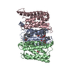
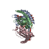
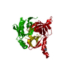
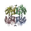
- PDB-5ys3: 1.8 angstrom crystal structure of Succinate-Acetate Permease from... -
+ Open data

- Basic information
| Entry | Database: PDB / ID: 5ys3 | ||||||||||||||||||
|---|---|---|---|---|---|---|---|---|---|---|---|---|---|---|---|---|---|---|---|
| Title | 1.8 angstrom crystal structure of Succinate-Acetate Permease from Citrobacter koseri | ||||||||||||||||||
 Components Components | Succinate-Acetate Permease | ||||||||||||||||||
 Keywords Keywords | TRANSPORT PROTEIN / organic anion channel / rectifying / unidirectional / dehydration | ||||||||||||||||||
| Function / homology |  Function and homology information Function and homology informationacetate:proton symporter activity / succinate transmembrane transport / plasma membrane Similarity search - Function | ||||||||||||||||||
| Biological species |  Citrobacter koseri (bacteria) Citrobacter koseri (bacteria) | ||||||||||||||||||
| Method |  X-RAY DIFFRACTION / SYNCHROTRON / SIRAS / Resolution: 1.823 Å X-RAY DIFFRACTION / SYNCHROTRON / SIRAS / Resolution: 1.823 Å | ||||||||||||||||||
 Authors Authors | Qiu, B. / Liao, J. | ||||||||||||||||||
| Funding support |  China, 5items China, 5items
| ||||||||||||||||||
 Citation Citation | Journal: Cell Res. / Year: 2018 Title: Succinate-acetate permease from Citrobacter koseri is an anion channel that unidirectionally translocates acetate Authors: Qiu, B. / Xia, B. / Zhou, Q. / Lu, Y. / He, M. / Hasegawa, K. / Ma, Z. / Zhang, F. / Gu, L. / Mao, Q. / Wang, F. / Zhao, S. / Gao, Z. / Liao, J. | ||||||||||||||||||
| History |
|
- Structure visualization
| Structure viewer | Molecule: MolmilJmol/JSmol |
|---|
- Downloads & links
-Download
| PDBx/mmCIF format | 5ys3.cif.gz | 125.2 KB | Display | PDBx/mmCIF format |
|---|---|---|---|---|
| PDB format | pdb5ys3.ent.gz | 98 KB | Display | PDB format |
| PDBx/mmJSON format | 5ys3.json.gz | Tree view | PDBx/mmJSON format | |
| Others |  Other downloads Other downloads |
-Validation report
| Arichive directory | https://data.pdbj.org/pub/pdb/validation_reports/ys/5ys3ftp://data.pdbj.org/pub/pdb/validation_reports/ys/5ys3 | HTTPS FTP |
|---|
-Related structure data


-Links
 PDBj
PDBj


- Assembly
| Deposited unit | 
| ||||||||
|---|---|---|---|---|---|---|---|---|---|
| 1 | 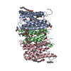
| ||||||||
| Unit cell |
|
-Components
| #1: Protein | Mass: 20473.240 Da / Num. of mol.: 3 Source method: isolated from a genetically manipulated source Source: (gene. exp.) Citrobacter koseri (strain ATCC BAA-895 / CDC 4225-83 / SGSC4696) (bacteria)Strain: ATCC BAA-895 / CDC 4225-83 / SGSC4696 / Gene: CKO_03375 / Production host: #2: Chemical | ChemComp-ACT /   Mass: 59.044 Da / Num. of mol.: 18 / Source method: obtained synthetically / Formula: C2H3O2 Mass: 59.044 Da / Num. of mol.: 18 / Source method: obtained synthetically / Formula: C2H3O2#3: Chemical | ChemComp-PLM /   Mass: 256.424 Da / Num. of mol.: 10 / Source method: obtained synthetically / Formula: C16H32O2 Mass: 256.424 Da / Num. of mol.: 10 / Source method: obtained synthetically / Formula: C16H32O2#4: Chemical | ChemComp-78M / ( 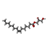  Mass: 314.460 Da / Num. of mol.: 7 / Source method: obtained synthetically / Formula: C18H34O4 Mass: 314.460 Da / Num. of mol.: 7 / Source method: obtained synthetically / Formula: C18H34O4#5: Water | ChemComp-HOH / |  Mass: 18.015 Da / Num. of mol.: 174 / Source method: isolated from a natural source / Formula: H2O Mass: 18.015 Da / Num. of mol.: 174 / Source method: isolated from a natural source / Formula: H2O |
|---|
-Experimental details
-Experiment
| Experiment | Method: X-RAY DIFFRACTION / Number of used crystals: 1 |
|---|
- Sample preparation
| Crystal | Density Matthews: 2.39 Å3/Da / Density % sol: 48.52 % |
|---|---|
| Crystal grow | Temperature: 298 K / Method: lipidic cubic phase / pH: 6.5 / Details: PEG 400, CaAc2, NaCl / PH range: 4.0-6.5 / Temp details: PEG 400 |
-Data collection
| Diffraction | Mean temperature: 100 K |
|---|---|
| Diffraction source | Source: SYNCHROTRON / Site: SPring-8  / Beamline: BL41XU / Wavelength: 0.979 Å / Beamline: BL41XU / Wavelength: 0.979 Å |
| Detector | Type: DECTRIS PILATUS3 S 6M / Detector: PIXEL / Date: Jun 16, 2015 |
| Radiation | Protocol: SINGLE WAVELENGTH / Monochromatic (M) / Laue (L): M / Scattering type: x-ray |
| Radiation wavelength | Wavelength: 0.979 Å / Relative weight: 1 |
| Reflection | Resolution: 1.8→50 Å / Num. obs: 51162 / % possible obs: 99.6 % / Redundancy: 6.9 % / Net I/σ(I): 17.5 |
| Reflection shell | Resolution: 1.8→1.86 Å / Redundancy: 6.6 % / Rmerge(I) obs: 0.8 / Mean I/σ(I) obs: 1.9 / CC1/2: 0.7 / % possible all: 97.6 |
- Processing
| Software |
| |||||||||||||||||||||||||||||||||||||||||||||||||||||||||||||||||||||||||||||||||||||||||||||||||||||||||||||||||||||||
|---|---|---|---|---|---|---|---|---|---|---|---|---|---|---|---|---|---|---|---|---|---|---|---|---|---|---|---|---|---|---|---|---|---|---|---|---|---|---|---|---|---|---|---|---|---|---|---|---|---|---|---|---|---|---|---|---|---|---|---|---|---|---|---|---|---|---|---|---|---|---|---|---|---|---|---|---|---|---|---|---|---|---|---|---|---|---|---|---|---|---|---|---|---|---|---|---|---|---|---|---|---|---|---|---|---|---|---|---|---|---|---|---|---|---|---|---|---|---|---|---|
| Refinement | Method to determine structure: SIRAS / Resolution: 1.823→39.728 Å / SU ML: 0.22 / Cross valid method: FREE R-VALUE / σ(F): 1.38 / Phase error: 19.37 / Stereochemistry target values: ML
| |||||||||||||||||||||||||||||||||||||||||||||||||||||||||||||||||||||||||||||||||||||||||||||||||||||||||||||||||||||||
| Solvent computation | Shrinkage radii: 0.9 Å / VDW probe radii: 1.11 Å / Solvent model: FLAT BULK SOLVENT MODEL | |||||||||||||||||||||||||||||||||||||||||||||||||||||||||||||||||||||||||||||||||||||||||||||||||||||||||||||||||||||||
| Refinement step | Cycle: LAST / Resolution: 1.823→39.728 Å
| |||||||||||||||||||||||||||||||||||||||||||||||||||||||||||||||||||||||||||||||||||||||||||||||||||||||||||||||||||||||
| Refine LS restraints |
| |||||||||||||||||||||||||||||||||||||||||||||||||||||||||||||||||||||||||||||||||||||||||||||||||||||||||||||||||||||||
| LS refinement shell |
|
