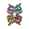
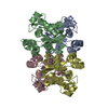
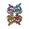
- PDB-5yih: Crystal structure of tetrameric Nucleoside diphosphate kinase at ... -
+
Open data
ID or keywords:
Loading...
-
Basic information
Entry
Database: PDB / ID: 5yih
Title
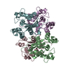
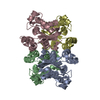
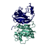
Crystal structure of tetrameric Nucleoside diphosphate kinase at 1.98 A resolution from Acinetobacter baumannii
Components
Nucleoside diphosphate kinase
Keywords
TRANSFERASE / Kinase
Function / homology
Function and homology information
nucleoside-diphosphate kinase / UTP biosynthetic process / CTP biosynthetic process / nucleoside diphosphate kinase activity / GTP biosynthetic process / ATP binding / metal ion binding / cytoplasm Similarity search - Function
Resolution: 1.98→68.8 Å / Cor.coef. Fo:Fc: 0.957 / Cor.coef. Fo:Fc free: 0.929 / SU B: 6.085 / SU ML: 0.164 / Cross valid method: THROUGHOUT / ESU R: 0.178 / ESU R Free: 0.17 / Details: HYDROGENS HAVE BEEN ADDED IN THE RIDING POSITIONS
Rfactor
Num. reflection
% reflection
Selection details
Rfree
0.25571
2259
4.9 %
RANDOM
Rwork
0.2021
-
-
-
obs
0.20471
43447
98.71 %
-
Solvent computation
Ion probe radii: 0.8 Å / Shrinkage radii: 0.8 Å / VDW probe radii: 1.2 Å
 Movie
Movie Controller

 Yorodumi
 Open data
 Basic information
 Components
 Keywords
 Function and homology information
 Acinetobacter baumannii (bacteria)
 X-RAY DIFFRACTION /
  Authors
 Citation
 Structure visualization
 Downloads & links
 Other downloads
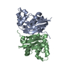

 PDBj
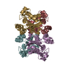
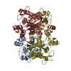
PDBj Assembly
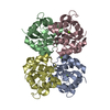

 Mass: 24.305 Da / Num. of mol.: 5 / Source method: obtained synthetically / Formula: Mg
 Mass: 18.015 Da / Num. of mol.: 343 / Source method: isolated from a natural source / Formula: H2O
 Sample preparation
 / Beamline: ID30B / Wavelength: 0.966 Å
 Processing