 Movie
Movie Controller

[English] 日本語
 Yorodumi
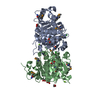
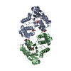
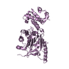
- PDB-5ybn: Fe(II)/(alpha)ketoglutarate-dependent dioxygenase PrhA in complex... -
+ Open data

- Basic information
| Entry | Database: PDB / ID: 5ybn | ||||||
|---|---|---|---|---|---|---|---|
| Title | Fe(II)/(alpha)ketoglutarate-dependent dioxygenase PrhA in complex with (alpha)ketoglutarate | ||||||
 Components Components | PrhA | ||||||
 Keywords Keywords | OXIDOREDUCTASE / alpha-kegoglutarate-dependent dioxygenase | ||||||
| Function / homology |  Function and homology information Function and homology informationparaherquonin biosynthetic process / Oxidoreductases; Acting on paired donors, with incorporation or reduction of molecular oxygen; With 2-oxoglutarate as one donor, and incorporation of one atom of oxygen into each donor / dioxygenase activity / metal ion binding Similarity search - Function | ||||||
| Biological species |  Penicillium brasilianum (fungus) Penicillium brasilianum (fungus) | ||||||
| Method |  X-RAY DIFFRACTION / SYNCHROTRON / MOLECULAR REPLACEMENT / Resolution: 2.104 Å X-RAY DIFFRACTION / SYNCHROTRON / MOLECULAR REPLACEMENT / Resolution: 2.104 Å | ||||||
 Authors Authors | Nakashima, Y. / Senda, M. | ||||||
 Citation Citation | Journal: Nat Commun / Year: 2018 Title: Structure function and engineering of multifunctional non-heme iron dependent oxygenases in fungal meroterpenoid biosynthesis. Authors: Nakashima, Y. / Mori, T. / Nakamura, H. / Awakawa, T. / Hoshino, S. / Senda, M. / Senda, T. / Abe, I. | ||||||
| History |
|
- Structure visualization
| Structure viewer | Molecule: MolmilJmol/JSmol |
|---|
- Downloads & links
-Download
| PDBx/mmCIF format | 5ybn.cif.gz | 134.8 KB | Display | PDBx/mmCIF format |
|---|---|---|---|---|
| PDB format | pdb5ybn.ent.gz | 102.3 KB | Display | PDB format |
| PDBx/mmJSON format | 5ybn.json.gz | Tree view | PDBx/mmJSON format | |
| Others |  Other downloads Other downloads |
-Validation report
| Arichive directory | https://data.pdbj.org/pub/pdb/validation_reports/yb/5ybnftp://data.pdbj.org/pub/pdb/validation_reports/yb/5ybn | HTTPS FTP |
|---|
-Related structure data
| Related structure data | 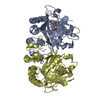 5yblC 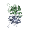 5ybmSC 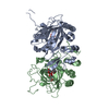 5yboC 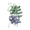 5ybpC 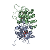 5ybqC 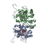 5ybrC 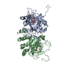 5ybsC 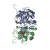 5ybtC S: Starting model for refinement C: citing same article ( |
|---|---|
| Similar structure data |


-Links
 PDBj
PDBj

- Assembly
| Deposited unit | 
| ||||||||
|---|---|---|---|---|---|---|---|---|---|
| 1 |
| ||||||||
| Unit cell |
|
-Components
| #1: Protein | Mass: 35166.781 Da / Num. of mol.: 2 / Fragment: UNP RESIDUES 6-294 Source method: isolated from a genetically manipulated source Source: (gene. exp.) Penicillium brasilianum (fungus) / Gene: prhA / Production host:  #2: Chemical |   Mass: 55.845 Da / Num. of mol.: 2 / Source method: obtained synthetically / Formula: Fe Mass: 55.845 Da / Num. of mol.: 2 / Source method: obtained synthetically / Formula: Fe#3: Chemical |   Mass: 146.098 Da / Num. of mol.: 2 / Source method: obtained synthetically / Formula: C5H6O5 Mass: 146.098 Da / Num. of mol.: 2 / Source method: obtained synthetically / Formula: C5H6O5#4: Water | ChemComp-HOH / |  Mass: 18.015 Da / Num. of mol.: 327 / Source method: isolated from a natural source / Formula: H2O Mass: 18.015 Da / Num. of mol.: 327 / Source method: isolated from a natural source / Formula: H2O |
|---|
-Experimental details
-Experiment
| Experiment | Method: X-RAY DIFFRACTION / Number of used crystals: 1 |
|---|
- Sample preparation
| Crystal | Density Matthews: 2.95 Å3/Da / Density % sol: 58.33 % |
|---|---|
| Crystal grow | Temperature: 293 K / Method: vapor diffusion, sitting drop / Details: PEG 3350, lithium citrate |
-Data collection
| Diffraction | Mean temperature: 95 K |
|---|---|
| Diffraction source | Source: SYNCHROTRON / Site: Photon Factory  / Beamline: BL-1A / Wavelength: 1.1 Å / Beamline: BL-1A / Wavelength: 1.1 Å |
| Detector | Type: DECTRIS EIGER X 4M / Detector: PIXEL / Date: Nov 4, 2016 |
| Radiation | Protocol: SINGLE WAVELENGTH / Monochromatic (M) / Laue (L): M / Scattering type: x-ray |
| Radiation wavelength | Wavelength: 1.1 Å / Relative weight: 1 |
| Reflection | Resolution: 2.1→43.1 Å / Num. obs: 44951 / % possible obs: 99.6 % / Redundancy: 3.5 % / Net I/σ(I): 17.1 |
| Reflection shell | Resolution: 2.1→2.2 Å |
- Processing
| Software |
| |||||||||||||||||||||||||||||||||||||||||||||||||||||||||||||||||||||||||||||||||||||||||||||||||||||||||
|---|---|---|---|---|---|---|---|---|---|---|---|---|---|---|---|---|---|---|---|---|---|---|---|---|---|---|---|---|---|---|---|---|---|---|---|---|---|---|---|---|---|---|---|---|---|---|---|---|---|---|---|---|---|---|---|---|---|---|---|---|---|---|---|---|---|---|---|---|---|---|---|---|---|---|---|---|---|---|---|---|---|---|---|---|---|---|---|---|---|---|---|---|---|---|---|---|---|---|---|---|---|---|---|---|---|---|
| Refinement | Method to determine structure: MOLECULAR REPLACEMENT Starting model: 5YBM Resolution: 2.104→43.087 Å / Cross valid method: FREE R-VALUE / Phase error: 24.52
| |||||||||||||||||||||||||||||||||||||||||||||||||||||||||||||||||||||||||||||||||||||||||||||||||||||||||
| Solvent computation | Shrinkage radii: 0.9 Å / VDW probe radii: 1.11 Å | |||||||||||||||||||||||||||||||||||||||||||||||||||||||||||||||||||||||||||||||||||||||||||||||||||||||||
| Refinement step | Cycle: LAST / Resolution: 2.104→43.087 Å
| |||||||||||||||||||||||||||||||||||||||||||||||||||||||||||||||||||||||||||||||||||||||||||||||||||||||||
| Refine LS restraints |
| |||||||||||||||||||||||||||||||||||||||||||||||||||||||||||||||||||||||||||||||||||||||||||||||||||||||||
| LS refinement shell |
|
