 Movie
Movie Controller

[English] 日本語
 Yorodumi
- PDB-5yb6: L-Amino acid oxidase/monooxygenase from Pseudomonas sp. AIU 813 -... -
+ Open data

- Basic information
| Entry | Database: PDB / ID: 5yb6 | ||||||
|---|---|---|---|---|---|---|---|
| Title | L-Amino acid oxidase/monooxygenase from Pseudomonas sp. AIU 813 - L-lysine complex | ||||||
 Components Components | L-amino acid oxidase/monooxygenase | ||||||
 Keywords Keywords | OXIDOREDUCTASE / L-amino acid oxidase/monooxygenase / flavin-containing monoamine oxidase family / flavin monooxygenases / L-lysine | ||||||
| Function / homology |  Function and homology information Function and homology informationtryptophan 2-monooxygenase activity / tryptophan 2-monooxygenase / auxin biosynthetic process / nucleotide binding Similarity search - Function | ||||||
| Biological species |  Pseudomonas sp. AIU 813 (bacteria) Pseudomonas sp. AIU 813 (bacteria) | ||||||
| Method |  X-RAY DIFFRACTION / SYNCHROTRON / MOLECULAR REPLACEMENT / molecular replacement / Resolution: 2.1 Å X-RAY DIFFRACTION / SYNCHROTRON / MOLECULAR REPLACEMENT / molecular replacement / Resolution: 2.1 Å | ||||||
 Authors Authors | Im, D. / Matsui, D. / Arakawa, T. / Isobe, K. / Asano, Y. / Fushinobu, S. | ||||||
 Citation Citation | Journal: FEBS Open Bio / Year: 2018 Title: Ligand complex structures of l-amino acid oxidase/monooxygenase from Authors: Im, D. / Matsui, D. / Arakawa, T. / Isobe, K. / Asano, Y. / Fushinobu, S. | ||||||
| History |
|
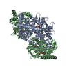
- Structure visualization
| Structure viewer | Molecule: MolmilJmol/JSmol |
|---|
- Downloads & links
-Download
| PDBx/mmCIF format | 5yb6.cif.gz | 465.4 KB | Display | PDBx/mmCIF format |
|---|---|---|---|---|
| PDB format | pdb5yb6.ent.gz | 375.6 KB | Display | PDB format |
| PDBx/mmJSON format | 5yb6.json.gz | Tree view | PDBx/mmJSON format | |
| Others |  Other downloads Other downloads |
-Validation report
| Arichive directory | https://data.pdbj.org/pub/pdb/validation_reports/yb/5yb6ftp://data.pdbj.org/pub/pdb/validation_reports/yb/5yb6 | HTTPS FTP |
|---|
-Related structure data
| Related structure data | 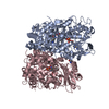 5yb7C  5yb8C  3we0S S: Starting model for refinement C: citing same article ( |
|---|---|
| Similar structure data |


-Links
 PDBj
PDBj

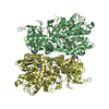
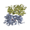
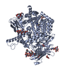
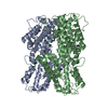
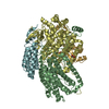
- Assembly
| Deposited unit | 
| |||||||||||||||||||||||||||||||||||||||||||||||||||||||||||||||||
|---|---|---|---|---|---|---|---|---|---|---|---|---|---|---|---|---|---|---|---|---|---|---|---|---|---|---|---|---|---|---|---|---|---|---|---|---|---|---|---|---|---|---|---|---|---|---|---|---|---|---|---|---|---|---|---|---|---|---|---|---|---|---|---|---|---|---|
| 1 | 
| |||||||||||||||||||||||||||||||||||||||||||||||||||||||||||||||||
| 2 | 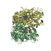
| |||||||||||||||||||||||||||||||||||||||||||||||||||||||||||||||||
| Unit cell |
| |||||||||||||||||||||||||||||||||||||||||||||||||||||||||||||||||
| Noncrystallographic symmetry (NCS) | NCS domain:
NCS domain segments:
NCS oper:
|
-Components
| #1: Protein | Mass: 64516.871 Da / Num. of mol.: 4 Source method: isolated from a genetically manipulated source Source: (gene. exp.) Pseudomonas sp. AIU 813 (bacteria) / Gene: laao, mog / Production host: #2: Chemical | ChemComp-FAD /   Mass: 785.550 Da / Num. of mol.: 4 / Source method: obtained synthetically / Formula: C27H33N9O15P2 / Comment: FAD*YM Mass: 785.550 Da / Num. of mol.: 4 / Source method: obtained synthetically / Formula: C27H33N9O15P2 / Comment: FAD*YM#3: Chemical | ChemComp-PG6 /   Mass: 266.331 Da / Num. of mol.: 4 / Source method: obtained synthetically / Formula: C12H26O6 Mass: 266.331 Da / Num. of mol.: 4 / Source method: obtained synthetically / Formula: C12H26O6#4: Chemical | ChemComp-LYS /   Type: L-peptide linking / Mass: 147.195 Da / Num. of mol.: 8 / Source method: obtained synthetically / Formula: C6H15N2O2 Type: L-peptide linking / Mass: 147.195 Da / Num. of mol.: 8 / Source method: obtained synthetically / Formula: C6H15N2O2#5: Water | ChemComp-HOH / |  Mass: 18.015 Da / Num. of mol.: 1026 / Source method: isolated from a natural source / Formula: H2O Mass: 18.015 Da / Num. of mol.: 1026 / Source method: isolated from a natural source / Formula: H2O |
|---|
-Experimental details
-Experiment
| Experiment | Method: X-RAY DIFFRACTION / Number of used crystals: 1 |
|---|
- Sample preparation
| Crystal | Density Matthews: 2.38 Å3/Da / Density % sol: 48.32 % |
|---|---|
| Crystal grow | Temperature: 293 K / Method: vapor diffusion, sitting drop / pH: 7 / Details: 20% PEG 3350, 0.15M DL-Malic acid |
-Data collection
| Diffraction | Mean temperature: 100 K | |||||||||||||||||||||||||||||||||||||||||||||||||||||||||||||||||||||||||||||||||||||||||||||||||||||||||||||||||||||||||||||||||||||||||||||||||||
|---|---|---|---|---|---|---|---|---|---|---|---|---|---|---|---|---|---|---|---|---|---|---|---|---|---|---|---|---|---|---|---|---|---|---|---|---|---|---|---|---|---|---|---|---|---|---|---|---|---|---|---|---|---|---|---|---|---|---|---|---|---|---|---|---|---|---|---|---|---|---|---|---|---|---|---|---|---|---|---|---|---|---|---|---|---|---|---|---|---|---|---|---|---|---|---|---|---|---|---|---|---|---|---|---|---|---|---|---|---|---|---|---|---|---|---|---|---|---|---|---|---|---|---|---|---|---|---|---|---|---|---|---|---|---|---|---|---|---|---|---|---|---|---|---|---|---|---|---|
| Diffraction source | Source: SYNCHROTRON / Site: Photon Factory  / Beamline: BL-5A / Wavelength: 1 Å / Beamline: BL-5A / Wavelength: 1 Å | |||||||||||||||||||||||||||||||||||||||||||||||||||||||||||||||||||||||||||||||||||||||||||||||||||||||||||||||||||||||||||||||||||||||||||||||||||
| Detector | Type: ADSC QUANTUM 315r / Detector: CCD / Date: Oct 29, 2012 | |||||||||||||||||||||||||||||||||||||||||||||||||||||||||||||||||||||||||||||||||||||||||||||||||||||||||||||||||||||||||||||||||||||||||||||||||||
| Radiation | Monochromator: Numerical link type Si(111) double crystal / Protocol: SINGLE WAVELENGTH / Monochromatic (M) / Laue (L): M / Scattering type: x-ray | |||||||||||||||||||||||||||||||||||||||||||||||||||||||||||||||||||||||||||||||||||||||||||||||||||||||||||||||||||||||||||||||||||||||||||||||||||
| Radiation wavelength | Wavelength: 1 Å / Relative weight: 1 | |||||||||||||||||||||||||||||||||||||||||||||||||||||||||||||||||||||||||||||||||||||||||||||||||||||||||||||||||||||||||||||||||||||||||||||||||||
| Reflection | Resolution: 2.1→50 Å / Num. obs: 139347 / % possible obs: 99.5 % / Redundancy: 3.4 % / Rmerge(I) obs: 0.126 / Χ2: 1.008 / Net I/σ(I): 4.4 | |||||||||||||||||||||||||||||||||||||||||||||||||||||||||||||||||||||||||||||||||||||||||||||||||||||||||||||||||||||||||||||||||||||||||||||||||||
| Reflection shell |
|
-Phasing
| Phasing | Method: molecular replacement |
|---|
- Processing
| Software |
| ||||||||||||||||||||||||||||||||||||||||||||||||||||||||||||
|---|---|---|---|---|---|---|---|---|---|---|---|---|---|---|---|---|---|---|---|---|---|---|---|---|---|---|---|---|---|---|---|---|---|---|---|---|---|---|---|---|---|---|---|---|---|---|---|---|---|---|---|---|---|---|---|---|---|---|---|---|---|
| Refinement | Method to determine structure: MOLECULAR REPLACEMENT Starting model: 3WE0 Resolution: 2.1→50 Å / Cor.coef. Fo:Fc: 0.911 / Cor.coef. Fo:Fc free: 0.866 / SU B: 6.978 / SU ML: 0.182 / Cross valid method: THROUGHOUT / σ(F): 0 / ESU R: 0.289 / ESU R Free: 0.229 Details: HYDROGENS HAVE BEEN ADDED IN THE RIDING POSITIONS U VALUES : REFINED INDIVIDUALLY
| ||||||||||||||||||||||||||||||||||||||||||||||||||||||||||||
| Solvent computation | Ion probe radii: 0.8 Å / Shrinkage radii: 0.8 Å / VDW probe radii: 1.2 Å | ||||||||||||||||||||||||||||||||||||||||||||||||||||||||||||
| Displacement parameters | Biso max: 97.09 Å2 / Biso mean: 19.388 Å2 / Biso min: 2.39 Å2
| ||||||||||||||||||||||||||||||||||||||||||||||||||||||||||||
| Refinement step | Cycle: final / Resolution: 2.1→50 Å
| ||||||||||||||||||||||||||||||||||||||||||||||||||||||||||||
| Refine LS restraints |
| ||||||||||||||||||||||||||||||||||||||||||||||||||||||||||||
| Refine LS restraints NCS | Ens-ID: 1 / Number: 8384 / Refine-ID: X-RAY DIFFRACTION
| ||||||||||||||||||||||||||||||||||||||||||||||||||||||||||||
| LS refinement shell | Resolution: 2.102→2.156 Å / Rfactor Rfree error: 0 / Total num. of bins used: 20
|
