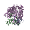
 Movie
Movie Controller


+ Open data

- Basic information
| Entry | Database: PDB / ID: 3we0 | ||||||
|---|---|---|---|---|---|---|---|
| Title | L-Amino acid oxidase/monooxygenase from Pseudomonas sp. AIU 813 | ||||||
 Components Components | L-amino acid oxidase/monooxygenase | ||||||
 Keywords Keywords | OXIDOREDUCTASE / Flavin-containing monoamine oxidase family / Rossmann Fold / Oxidoreductase (oxidase and monooxygenase) | ||||||
| Function / homology |  Function and homology information Function and homology informationtryptophan 2-monooxygenase activity / tryptophan 2-monooxygenase / auxin biosynthetic process / nucleotide binding Similarity search - Function | ||||||
| Biological species |  Pseudomonas sp. (bacteria) Pseudomonas sp. (bacteria) | ||||||
| Method |  X-RAY DIFFRACTION / SYNCHROTRON / SAD / Resolution: 1.9 Å X-RAY DIFFRACTION / SYNCHROTRON / SAD / Resolution: 1.9 Å | ||||||
 Authors Authors | Im, D.H. / Matsui, D. / Fukuta, Y. / Fushinobu, S. / Isobe, K. / Asano, Y. | ||||||
 Citation Citation | Journal: FEBS Open Bio / Year: 2014 Title: Mutational and crystallographic analysis of l-amino acid oxidase/monooxygenase from Pseudomonas sp. AIU 813: Interconversion between oxidase and monooxygenase activities Authors: Matsui, D. / Im, D.H. / Sugawara, A. / Fukuta, Y. / Fushinobu, S. / Isobe, K. / Asano, Y. #1: Journal: J. Biosci. Bioeng. / Year: 2012Title: Purification and characterization of an L-amino acid oxidase from Pseudomonas sp. AIU 813 Authors: Isobe, K. / Sugawara, A. / Domon, H. / Fukuta, Y. / Asano, Y. | ||||||
| History |
|
- Structure visualization
| Structure viewer | Molecule: MolmilJmol/JSmol |
|---|
- Downloads & links
-Download
| PDBx/mmCIF format | 3we0.cif.gz | 231.4 KB | Display | PDBx/mmCIF format |
|---|---|---|---|---|
| PDB format | pdb3we0.ent.gz | 183.7 KB | Display | PDB format |
| PDBx/mmJSON format | 3we0.json.gz | Tree view | PDBx/mmJSON format | |
| Others |  Other downloads Other downloads |
-Validation report
| Arichive directory | https://data.pdbj.org/pub/pdb/validation_reports/we/3we0ftp://data.pdbj.org/pub/pdb/validation_reports/we/3we0 | HTTPS FTP |
|---|
-Related structure data
| Similar structure data |
|---|


-Links
 PDBj
PDBj
- Assembly
| Deposited unit | 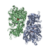
| ||||||||
|---|---|---|---|---|---|---|---|---|---|
| 1 | 
| ||||||||
| Unit cell |
|
-Components
| #1: Protein | Mass: 64516.871 Da / Num. of mol.: 2 Source method: isolated from a genetically manipulated source Source: (gene. exp.) Pseudomonas sp. (bacteria) / Strain: AIU 813 / Gene: laao / Plasmid: pET15b-laao / Production host: #2: Chemical |   Mass: 785.550 Da / Num. of mol.: 2 / Source method: obtained synthetically / Formula: C27H33N9O15P2 / Comment: FAD*YM Mass: 785.550 Da / Num. of mol.: 2 / Source method: obtained synthetically / Formula: C27H33N9O15P2 / Comment: FAD*YM#3: Water | ChemComp-HOH / |  Mass: 18.015 Da / Num. of mol.: 404 / Source method: isolated from a natural source / Formula: H2O Mass: 18.015 Da / Num. of mol.: 404 / Source method: isolated from a natural source / Formula: H2OSequence details | AUTHORS STATE THAT THE GENEBANK ACCESSION NUMBER FOR THIS SEQUENCE IS AB830473. | |
|---|
-Experimental details
-Experiment
| Experiment | Method: X-RAY DIFFRACTION / Number of used crystals: 1 |
|---|
- Sample preparation
| Crystal | Density Matthews: 2.47 Å3/Da / Density % sol: 50.29 % |
|---|---|
| Crystal grow | Temperature: 293.15 K / Method: vapor diffusion, sitting drop / pH: 7.6 Details: 8% polyethylene glycol (PEG) 4000, 0.1M Na-acetate(pH4.6), pH 7.6, VAPOR DIFFUSION, SITTING DROP, temperature 293.15K |
-Data collection
| Diffraction | Mean temperature: 100 K |
|---|---|
| Diffraction source | Source: SYNCHROTRON / Site: Photon Factory  / Beamline: BL-1A / Wavelength: 1 Å / Beamline: BL-1A / Wavelength: 1 Å |
| Detector | Type: ADSC QUANTUM 270 / Detector: CCD / Date: Nov 13, 2011 |
| Radiation | Monochromator: Cryo-cooled channel-cut Si(111) / Protocol: SINGLE WAVELENGTH / Monochromatic (M) / Laue (L): M / Scattering type: x-ray |
| Radiation wavelength | Wavelength: 1 Å / Relative weight: 1 |
| Reflection | Resolution: 1.9→50 Å / Num. all: 101012 / Num. obs: 98486 / % possible obs: 97.5 % / Observed criterion σ(I): -3 / Redundancy: 6.3 % / Biso Wilson estimate: 23.3 Å2 / Rsym value: 0.082 / Net I/σ(I): 24.7 |
| Reflection shell | Resolution: 1.9→1.93 Å / Redundancy: 6.2 % / Mean I/σ(I) obs: 3.2 / Num. unique all: 4930 / Rsym value: 0.507 / % possible all: 99.9 |
- Processing
| Software |
| |||||||||||||||||||||||||
|---|---|---|---|---|---|---|---|---|---|---|---|---|---|---|---|---|---|---|---|---|---|---|---|---|---|---|
| Refinement | Method to determine structure: SAD / Resolution: 1.9→30.38 Å / Cor.coef. Fo:Fc: 0.945 / Cor.coef. Fo:Fc free: 0.929 / SU B: 0.002 / SU ML: 0 / Cross valid method: THROUGHOUT / ESU R: 0.127 / ESU R Free: 0.154 / Stereochemistry target values: MAXIMUM LIKELIHOOD
| |||||||||||||||||||||||||
| Solvent computation | Ion probe radii: 0.8 Å / Shrinkage radii: 0.8 Å / VDW probe radii: 1.2 Å / Solvent model: MASK | |||||||||||||||||||||||||
| Displacement parameters | Biso mean: 28.961 Å2
| |||||||||||||||||||||||||
| Refine analyze |
| |||||||||||||||||||||||||
| Refinement step | Cycle: LAST / Resolution: 1.9→30.38 Å
| |||||||||||||||||||||||||
| Refine LS restraints |
| |||||||||||||||||||||||||
| LS refinement shell | Resolution: 1.9→1.951 Å / Total num. of bins used: 20
|
